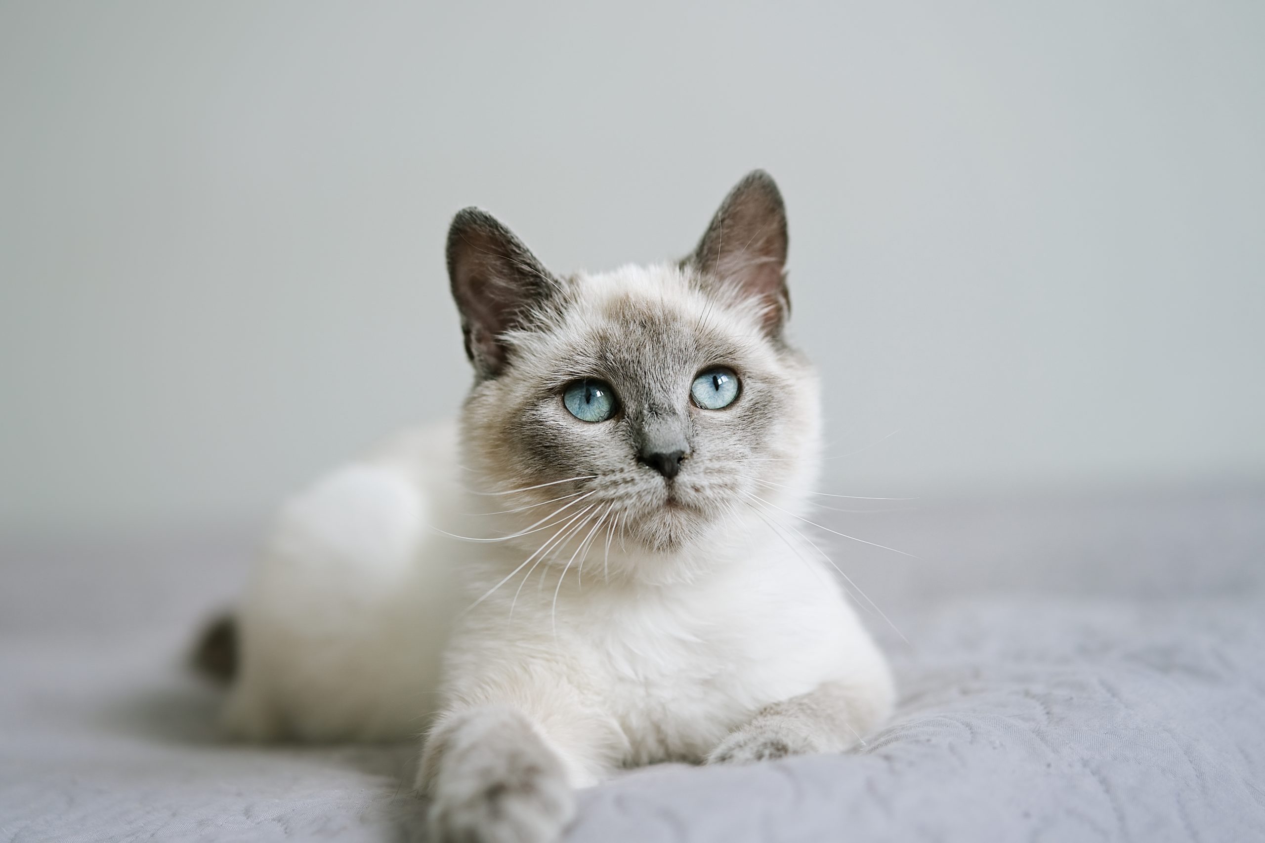
Mickey
 Case Background
Name: Mickey
Age: 14 years
Sex: Male
Breed: Feline, Domestic Medium Hair
Weight: 6.2 kg
Reason for visit: Murmur evaluation, heard by general veterinarian at wellness appointment.
Age: 14 years
Sex: Male
Breed: Feline, Domestic Medium Hair
Weight: 6.2 kg
Reason for visit: Murmur evaluation, heard by general veterinarian at wellness appointment.
 Clinical History
Please review Mickey’ clinical history.
Attitude/demeanor: Calm, alert
Coughing: No cough reported
Respirations: Eupneic, 32 breaths per minute
Exercise tolerance: No change reported
Sleep patterns: Sleeping well, most of the day
Weight change (loss or gain): No change in weight
Appetite: Normal appetite
Usual diet: Iams® Feline Senior
Vomiting: Rare vomiting, 1-2x monthly
Diarrhea: None noted
Syncope: None observed
Change in urinary habits: None observed
Change in drinking habits: None observed
 Physical Exam - General
Please review the results of Mickey’s physical exam.
Body condition: Slightly over-conditioned, BCS 6/9
Attitude: Calm, alert
Mobility | gait: Not assessed
Posture: Normal
Hydration: Normal
Body temperature: 102.1 F
Arterial pulse – rate, regularity, intensity: 176 beats/min, regular, slightly hyperkinetic
Respiratory effort: Effort 32 breaths per minute, normal
Mucous membranes – color & CRT: Pink
Jugular venous pulse & pressure: Slight distension, no pulsation
Abdominal palpatation: Both kidneys have irregular contours on palpation; the rest of the abdominal palpation is non-tender and normal
Lymph nodes: Normal
Oral cavity: Mild gingivitis and plaque
Other abnormalities: Slightly rough, dry hair coat
 Physical Exam - Auscultation
Let’s auscult Mickey’s heart & lungs. (Recommend listening with high-end headphones)
How do you interpret Mickey's cardiac auscultation (recorded at the left parasternal border)?
 Physical Exam - Differential Diagnosis
The following are potential diagnoses for you to consider at this time. Based on the history and the physical examination, please indicate the likelihood of each as:
- High (could explain most or all of the signs)
- Possible (less likely to explain most of the signs)
- Unlikely
Functional murmur
Hypertrophic cardiomyopathy
Degenerative mitral valve disease
Hyperthyroidism
Hypertensive heart disease
Diagnostic Test Selection
Non-invasive blood pressure
CBC with platelet count
Serum biochemical profile (includes electrolytes)
Urinalysis
Serum thyroxine (T4)
NT-ProBNP
Cardiac troponin-I
Thoracocentesis or abdominocentesis for diagnosis or therapy
Thoracic radiographs
Abdominal radiographs
Echocardiogram doppler studies
Abdominal ultrasound
ECG rhythm strip or 6 lead ECG
 Blood Pressure
Systolic blood pressure: 142 mmHg
Diastolic blood pressure: Not available for this case
Mean blood pressure: Not available for this case Consensus Statements of the American College of Veterinary Internal Medicine (ACVIM) provides the veterinary community with up-to-date information on the pathophysiology, diagnosis, and treatment of clinically important animal diseases. In 2018, ACVIM published updated guidelines for the identification, evaluation, and management of systemic hypertension in dogs and cats in the Journal of Veterinary Internal Medicine.
Click here to view and download a PDF of the ACVIM Consensus Statement, guidelines for the identification, evaluation, and management of systemic hypertension in dogs and cats.
 Radiography
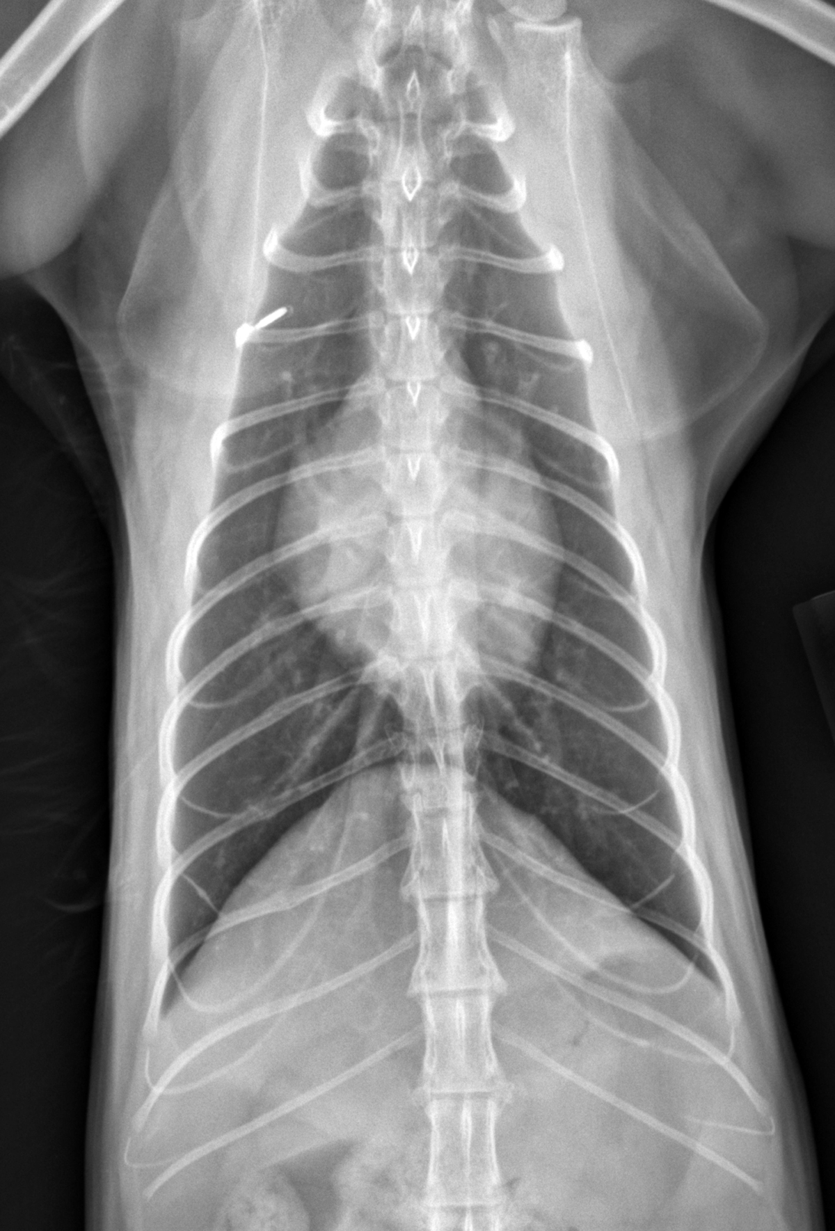
Please review Mickey’s thoracic radiographs.
Click here for Mickey’s radiograph viewer (measure VHS here) View the ventral dorsal radiograph
Click here for ventral dorsal view
Click here for lateral view
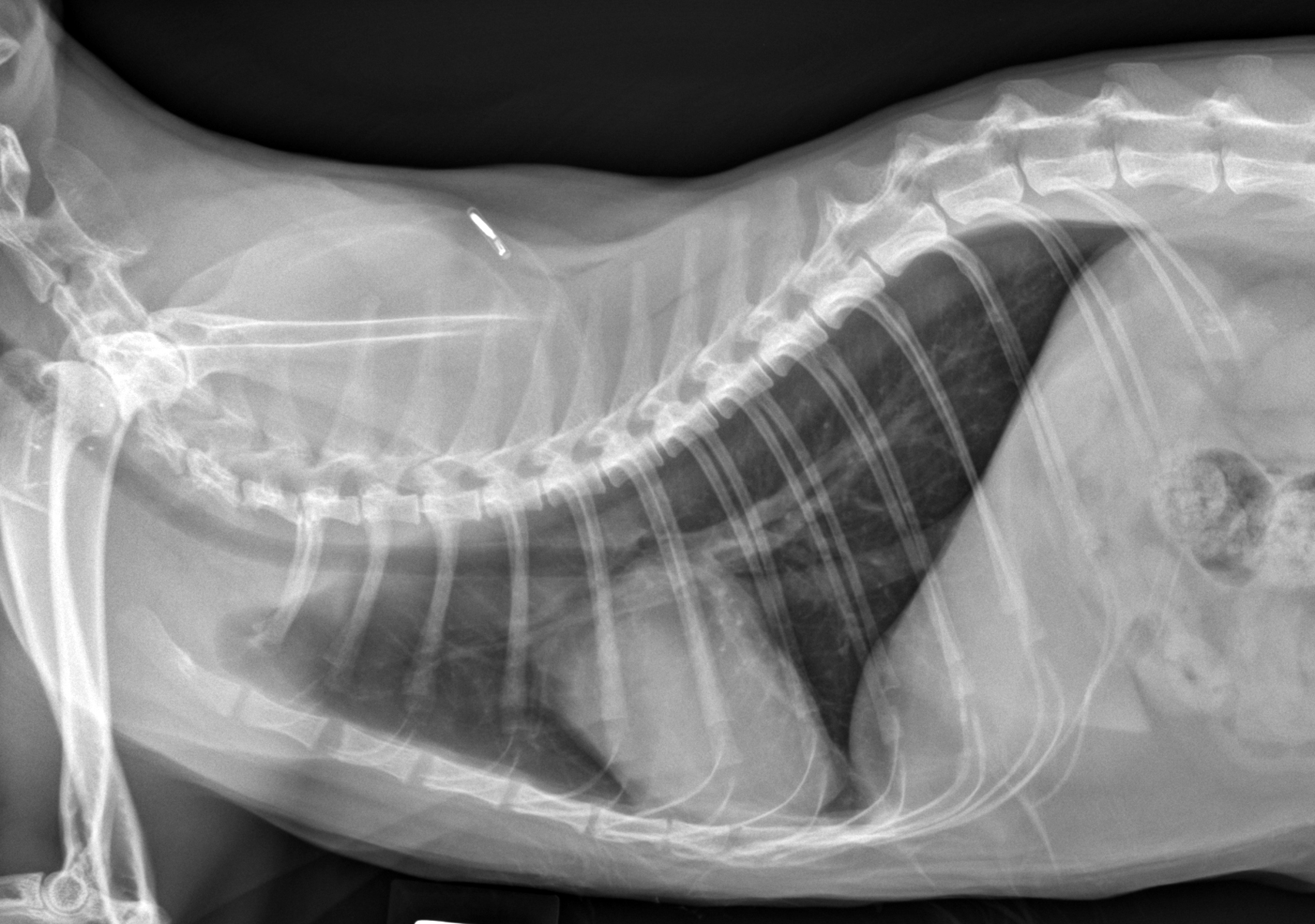
What is the vertebral heart size (VHS)?
Is Mickey's heart enlarged?
Is there evidence of congestive heart failure present (pleural effusion or pulmonary edema)?
 Clinical Labs
Serum chemistries
BUN: 34 mg/dL, Normal: 5 – 20 mg/dL
Creatinine: 2.2 mg/dL, Normal: 0.9 – 2.1 mg/dL
Sodium: 144 mEq/L, Normal:146 – 156 mEq/L
Potassium: 3.8 mEq/L, Normal: 3.2 – 5.5 mEq/L
Chloride: 118 mEq/L, Normal: 114 – 126 mEq/L
ALT: 52 IU/L, Normal: 20 – 95 IU/L
ALP: 80 IU/L, Normal: 15 – 65 IU/L
Heartworm
Heartworm: Not performed
Urinalysis
Urinalysis – USG: 1.018
Urinalysis – protein: Trace
Urinalysis – biochemical: No significant findings
Urinalysis – sediment evaluation: No significant findings
CBC
White blood cells: Not evaluated
Red blood cells: Normal PCV – 34%
Platelets: Not Evaluated
Total T4
Total T4 test results: 2.1 micrograms/dL
NT-proBNP
NT-proBNP test results: 497 pmol/L
 Echocardiography
Your technician just returned with the results of Mickey’s echo.
Please view the video and interpretations that follow.
Click here to watch Mickey's echo
LV chamber size and thickness: The left venticular walls show symmetrical concentric hypertrophy, measuring thicker than normal. Normal cats have walls that measure 3 to 5 mm in end-diastole with any measurement over 6 mm consistent with hypertrophy. Mickey’s left ventricular freewall and septum measure from 6.5 to 8.5 mm in thickness. In addition, his papillary muscles, seen best in the short-axis images, are severely hypertrophied.
Left atrial size: Left atrial size appears mildly dilated, but measurements are required to confirm enlargement. His left atrium did measure slightly larger than normal at 17 mm at end-systole (normal cats have a left atrium that typically measures 11 to 16 mm in diameter).
LVIDd & LVIDs: Left ventricular internal dimensions appear normal to slightly reduced as a consequence of the concentric hypertrophy.
LV shortening fraction: Mickey’s fractional shortening is normal to hyperdynamic. Most cats have a fractional shortening of 35 to 55%; Mickey’s measured 57% likely related to the thick walls compromising his systolic dimension.
RA, RV and pulmonary artery: Images of the right heart are not fully shown, but were normal in this cat.
Effusions: There are no effusions apparent.
Doppler results: Color Doppler shows turbulence in the left ventricular outflow tract beginning at the site of mitral-septal contact. This is a consequence of altered left ventricular, papillary, and mitral valve geometry resulting in SAM – systolic anterior motion of the mitral valve.
 Referral
The echocardiogram shown in this case study was acquired at the cardiologist, following referral from Mickey’s general veterinarian. The blood work, blood pressure, and thoracic radiographs were all obtained by the general veterinarian prior to referral.
 Diagnosis & Treatment
You’re ready to form a diagnosis and treatment plan for Mickey! Please select your answer to each question below.
What is your diagnosis for Mickey?
What treatment(s) would you recommend for Mickey?
Follow-up Treatment
Follow-up treatment: Follow-up is recommended with Mickey’s general veterinarian in 7-10 days to evaluate the response to the atenolol. In general, the response to atenolol is evaluated by monitoring the cat’s heart rate and blood pressure while receiving the medication as well as an assessment for any clinical signs. An excess of beta-blockade would manifest as a lethargic and less active, or even syncopal, cat. On examination of such a cat on too much atenolol, the heart rate is below 140bpm even during stress and blood pressure may be low. Under-dosing of the atenolol is suspected based on an in-hospital heart rate of >170 bpm. In general, a cat on an optimal dose of atenolol will have no change in clinical history and a heart rate in the hospital (e.g. under stress) of 140 to 160bpm. Doses are adjusted to achieve this finding. Once on a stable and appropriate dose of atenolol, a recheck echocardiogram is advised in 3-4 months and then once to twice yearly thereafter to monitor for progressive cardiac enlargement.
 Post Test - CE
To qualify for CE credit, please answer the following 5 questions.
THORACIC RADIOGRAPHS CAN PROVIDE A DIAGNOSIS OF HEART DISEASE WITH HIGH SENSITIVITY AND SPECIFICITY IN THE CAT.
WHICH OF THE FOLLOWING IS CORRECT, REGARDING AN ELEVATED NT-PROBNP (ABOVE 100 PMOL/L) IN A CAT?
SYSTOLIC ANTERIOR MOTION OF THE MITRAL VALVE (OR SAM) IN THE SETTING OF HYPERTROPHIC CARDIOMYOPATHY IMPLIES WHICH OF THE FOLLOWING IS TRUE?
THERAPY FOR THE ASYMPTOMATIC CAT WITH HYPERTROPHIC CARDIOMYOPATHY (HCM) SHOULD INCLUDE WHICH OF THE FOLLOWING?
FOLLOW-UP FOR THE CAT RECEIVING ATENOLOL FOR THE TREATMENT OF HYPERTROPHIC OBSTRUCTIVE CARDIOMYOPATHY SHOULD INCLUDE WHICH OF THE FOLLOWING?
 RACE Certification
RACE Certification
Fill out the following form in order to receive your certificate.