Poppy
 Case Background
Name: Poppy
Age: 16 years
Sex: Female, spayed
Breed: Feline, domestic medium hair
Age: 16 years
Sex: Female, spayed
Breed: Feline, domestic medium hair
 Clinical History
Please review Poppy’s clinical history.
Seen on emergency service with 6 day history of increased respiratory effort and syncopal episode tonight. Physical exam revealed a bright and alert patient with a gallop rhythm (HR 160 bpm), a 2/6 left sternal border murmur and bilateral pulmonary crackles. A freely movable soft subcutanous mass was palpated over the dorsal spine.
 Radiographs
View Poppy's left lateral radiograph
 View Poppy's annotated left lateral radiograph
 View Poppy's ventrodorsal radiograph
 View Poppy's annotated ventrodorsal radiograph
 Click here for Poppy’s radiograph viewer (measure VHS)
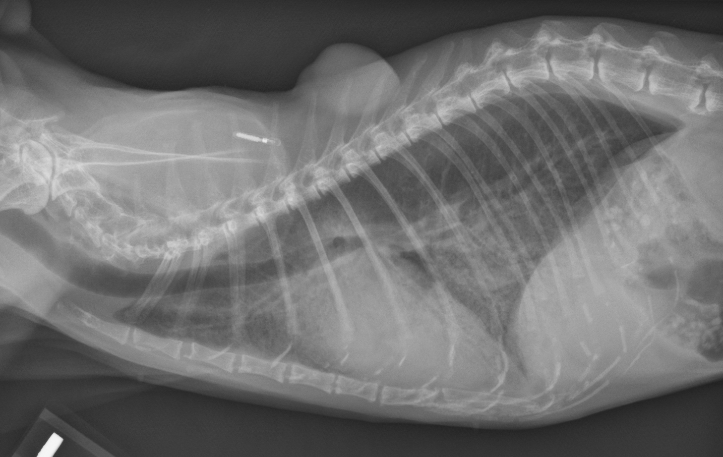
Poppy's left lateral radiograph
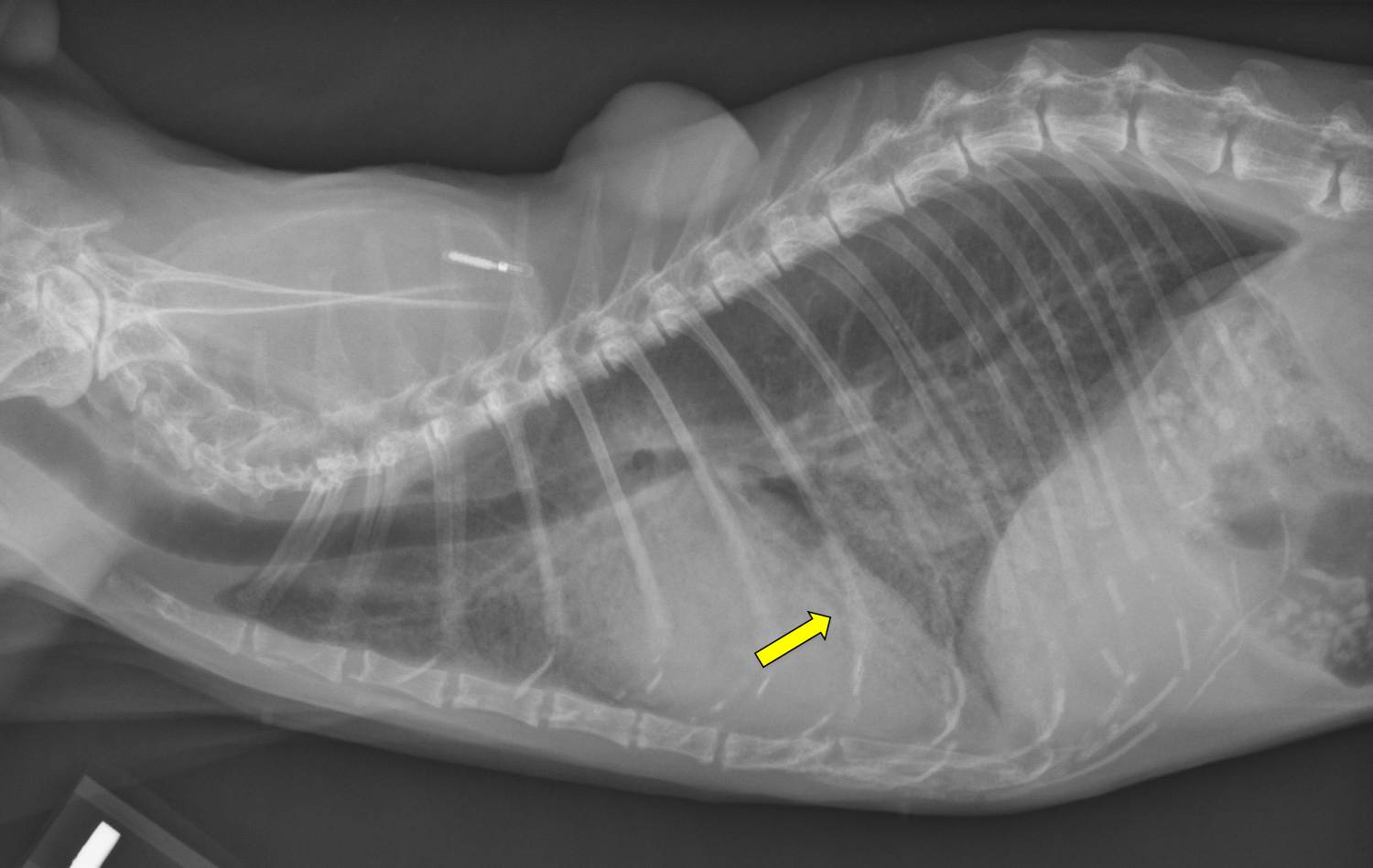
Poppy's annotated left lateral radiograph
Poppy's ventrodorsal radiograph
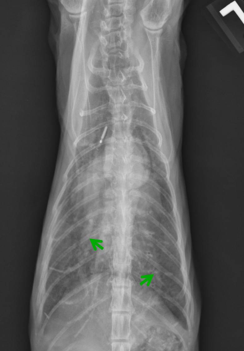
Poppy's annotated ventrodorsal radiograph
Technical details
Location of images: Thoracic radiographs obtained.
Views of images: Left lateral and ventrodorsal (VD) views.Radiographic findings
Technical issues: Good exposure, VD view is slightly twisted. Subcutaneous microchip visible. Dorsal subcutaneous mass visible on lateral view.
Cardiac size including VHS: VHS 9.5, moderate to severe generalized cardiomegaly.
Other findings: Diffuse moderate to severe alveolar infiltrates distributed ventrally in cranial and caudal lung fields bilaterally. The caudal pulmonary arteries appear to be enlarged (green arrows). Air bronchograms are visible in the right middle lung lobe on the lateral view (yellow arrow).
 Diagnosis & Treatment
Radiographic interpretation: Probable congestive heart failure (pulmonary edema).
Discussion: Cardiac enlargement combined with moderate to severe alveolar infiltrates and physical findings of a gallop rhythm and left sided heart murmur are strongly suggestive of congestive heart failure in this patient. Cats frequently develop pulmonary hypertension when left-sided heart failure is present; in this cat, enlarged pulmonary arteries are seen on the VD view, consistent with increased pulmonary arterial pressures. In this case, the underlying heart disease is hypertrophic cardiomyopathy (based on echocardiogram). It is important to note that the underlying heart disease causing the heart failure cannot typically be diagnosed by radiographic appearance.
Treatment/management: Congestive heart failure with pulmonary edema in cats can be treated with provision of additional oxygen (in this case, an oxygen cage) and injectable furosemide. In most cats, beginning doses of furosemide of 2 mg/kg IM administered every 1-3 times over the first 12 hours will result in clinical improvement, often noted as a decrease in respiratory rate in the resting animal.