
Shivers
 Case Background
Name: Shivers
Age: 11 years
Sex: Male, neutered
Breed: Miniature Poodle
Age: 11 years
Sex: Male, neutered
Breed: Miniature Poodle
 Clinical History
Please review Shivers’ clinical history
This dog had a primary complaint of rapidly progressive coughing (began 2 weeks ago, but is much more severe over the past 48 hours) accompanied by lethargy and increased respiratory effort. Has a history of a heart murmur, first detected 2 years ago.
 Radiographs
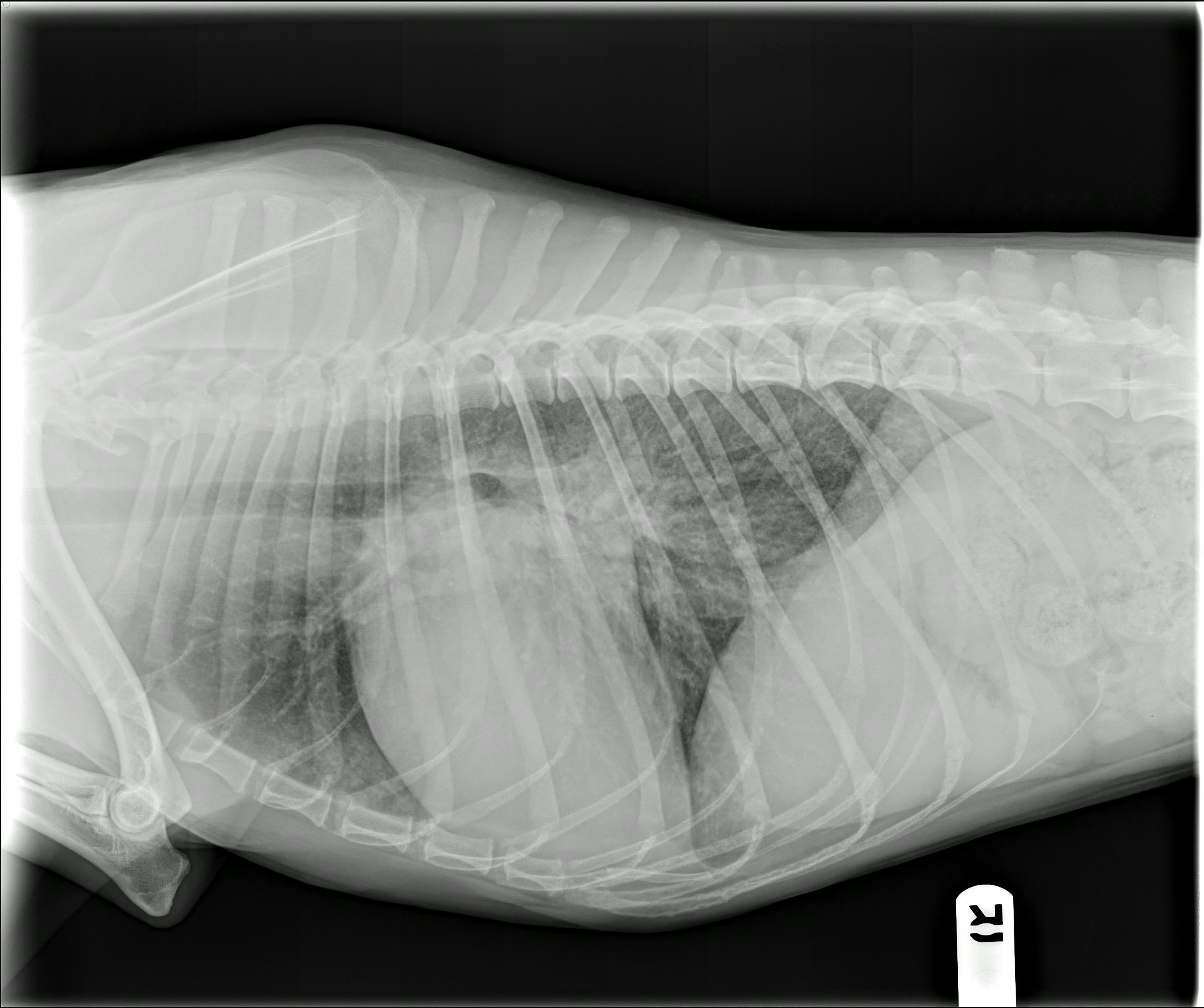
View Shivers' right lateral radiograph
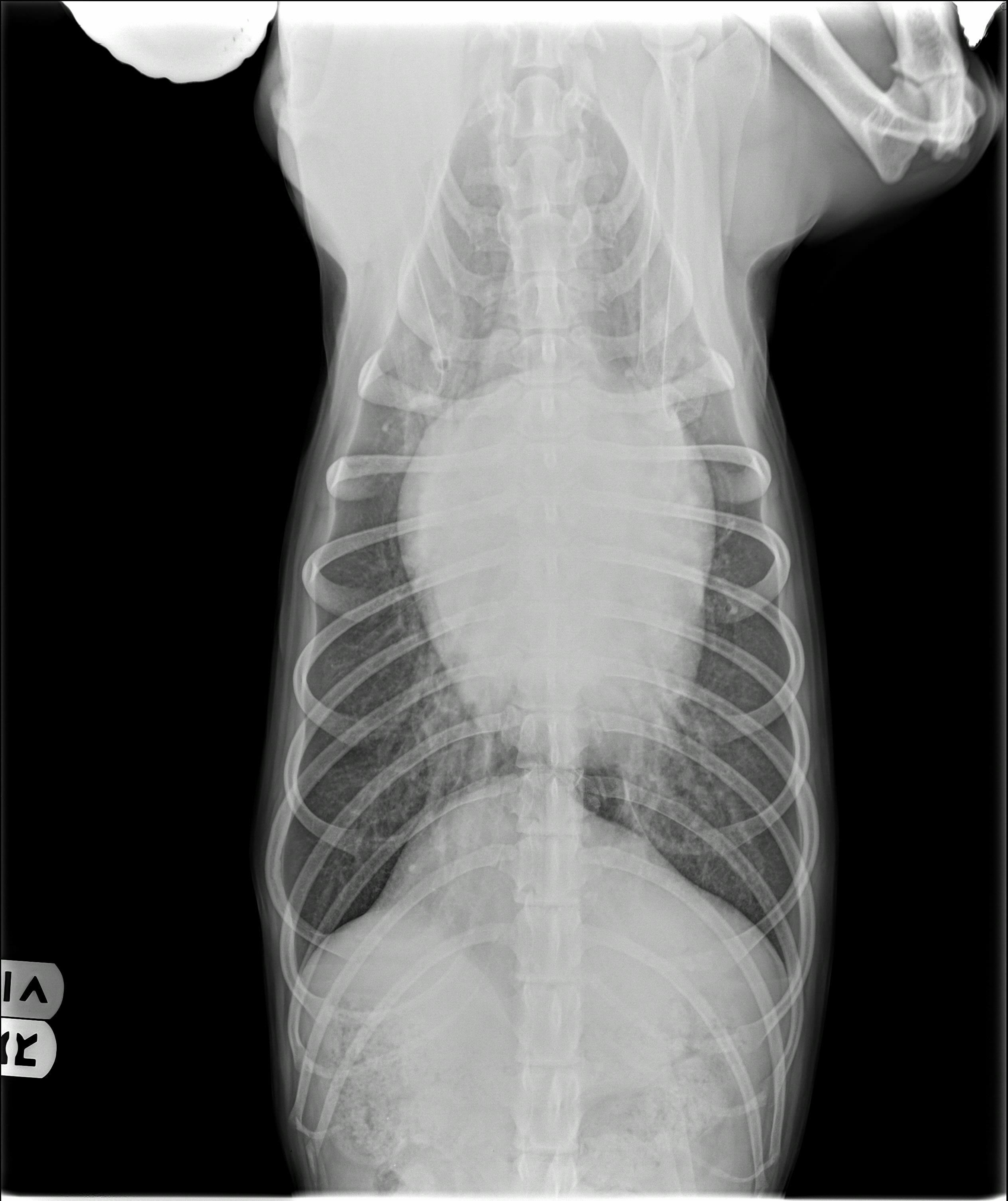
 View Shivers' ventrodorsal radiograph
Shivers' right lateral radiograph
Shivers' ventrodorsal radiograph
Click here for Shivers’ radiograph viewer (measure VHS and VLAS)
Radiographic findings
The technical quality is good, though it could be collimated to exclude the abdomen. The cardiac silhouette is enlarged with a VHS of 12.4 and a VLAS of 2.7. There is severe left-sided heart enlargement with loss of the caudal waist, indicating left atrial dilation. On the VD view, the cardiac silhouette appears to be wide and somewhat elongated, with a broad left auricular bulge at the 2-3 o’clock position on the cardiac silhouette, consistent with left atrial chamber enlargement. There is a caudal interstitial pattern that is located toward the midline and radiates outward toward the thoracic wall, with the heaviest radiographic density overlaying the left apex of the cardiac silhouette. The pulmonary veins are distended relative to the corresponding arteries.
 Diagnosis & Treatment
Radiographic interpretation: Left-sided Congestive Heart Failure (pulmonary edema) likely secondary to degenerative valve disease.
Discussion: The enlarged heart, venous distention and distribution of the interstitial pattern are all consistent with congestive heart failure. The murmur and breed suggest degenerative valve disease as the most likely cause of the heart enlargement and pulmonary venous congestion. An echocardiogram was performed and revealed moderate to severe left atrial dilation with moderate left ventricular dilation with severe thickening and prolapse of the anterior leaflet of the mitral valve. Hyperdynamic left ventricular systolic function was found, secondary to mitral regurgitation. There was a large jet of mitral regurgitation with an elevated early diastolic E wave velocity (mitral inflow Doppler study) consistent with elevated atrial pressure. Mild tricuspid regurgitation was also present, with a high normal regurgitant velocity consistent with a mild elevation in pulmonary arterial pressure (42mmHg pressure gradient). There were normal aortic and pulmonary flow velocities. No pericardial effusion present.
Treatment/management: The patient was given a furosemide injection (30mg) subcutaneously and sent home on: Furosemide 20mg tablet every 8 hours by mouth for 3 days then decreased to every 12 hours. Enalapril 5mg tablet every 12 hours by mouth. Pimobendan 2.5mg every 12 hours by mouth. He returned in 7 days for a renal panel and repeat radiographs (see follow up films) which showed improvement in the interstitial pattern, but the pulmonary venous distention had not resolved. Spironolactone 12.5mg every 12 hours was added at that visit.
 Follow-Up
Please view Shivers’ re-check radiographs
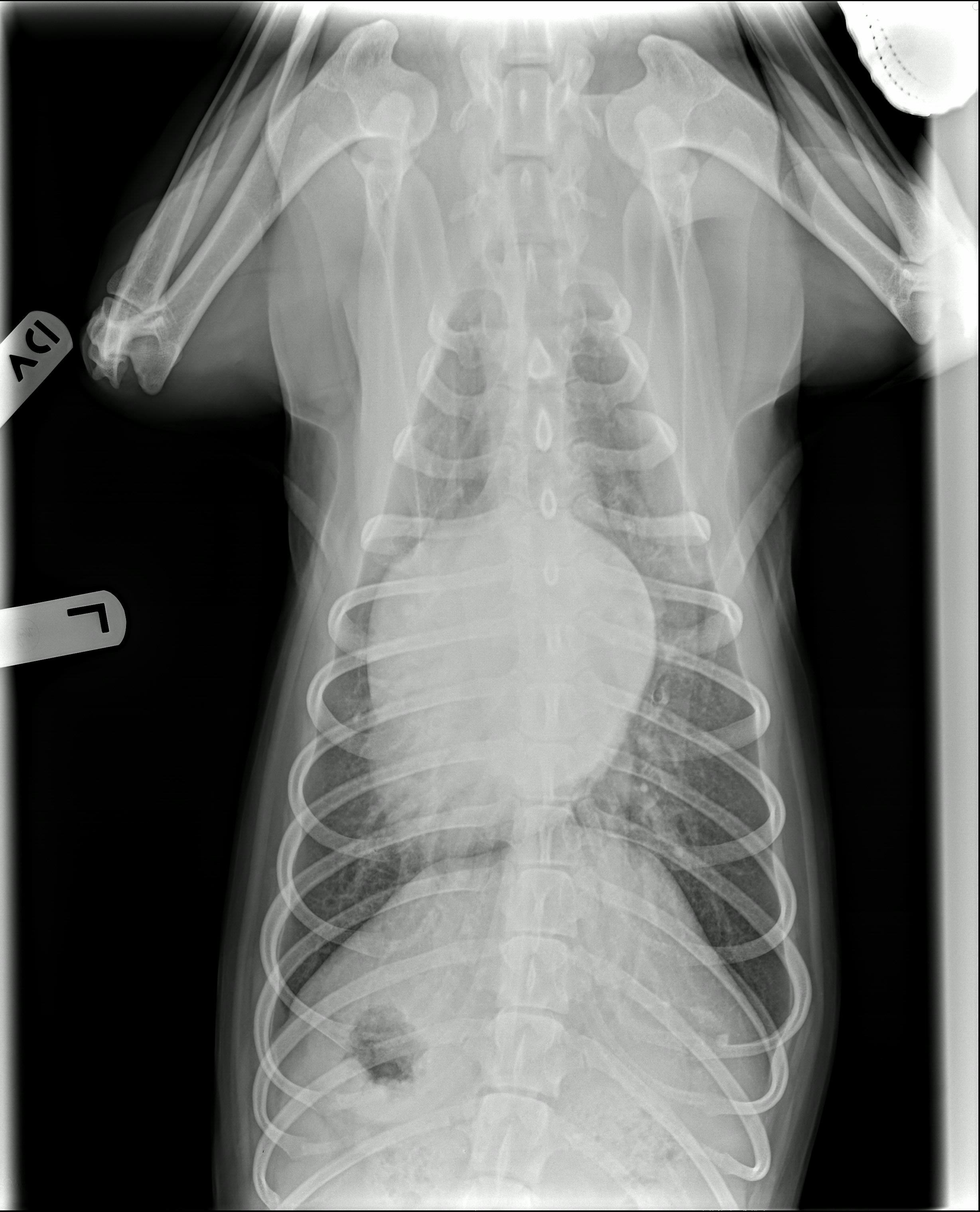
 View Shiver's ventrodorsal recheck radiograph
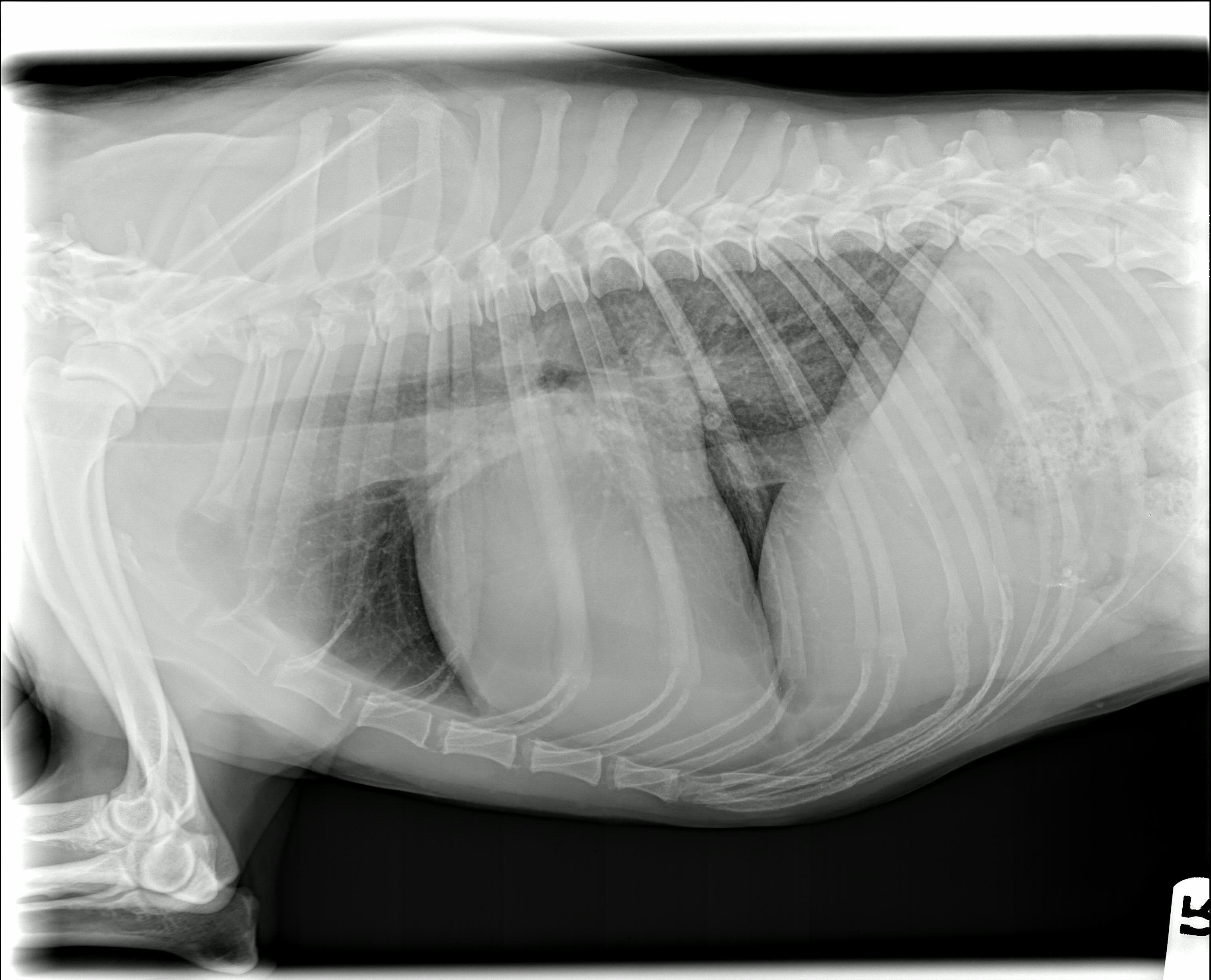
View Shiver's right lateral recheck radiograph
Shiver's right lateral recheck radiograph
Shiver's ventrodorsal recheck radiograph