Hailey
 Case Background
Age: 7 years
Sex: Female, spayed
Breed: Cavalier King Charles Spaniel
Weight: 26.4 pounds (12 kg)
Reason for Visit: Hailey was presented for her yearly physical examination. The family has reported no clinical signs other than bad breath. There has been no cough, respiratory distress, weakness, decreased exercise tolerance or change in appetite
Medications: Heartgard® Plus, no other chronic medications. Last heartworm test was about 2 years prior to this exam.
 Clinical History
Please review Hailey’s clinical history.
Attitude/Demeanor: Normal
Coughing: No cough
Abnormal Respirations: None
Exercise intolerance: None
Sleep Patterns: Unchanged
Weight Change (loss or gain): None
Appetite: No change
Usual Diet: Eukanuba™ Adult with some “people food”
Vomiting: None
Diarrhea: None
Syncope: None
Change in urinary habits: None
Change in drinking habits: None
Other symptoms or signs: None
 Physical Exam - General
Please review the results of Hailey’s physical examination.
Body Condition: Mildly overweight – BCS 6/9
Attitude: Alert, calm
Mobility | Gait: Normal gait
Posture: Laying in sternal recumbency, resting comfortably
Hydration: Normal
Body Temperature: 100.6 F
Arterial pulse – rate, regularity, intensity: 120 bpm, regularly irregular (with ventilation), normal strength
Rate & Respiratory: Normal effort, 22 breaths per minute
Mucous Membranes – Color & CRT: Pink, <2sec
Jugular Venous Pulse & Pressure: Normal
Abdominal Palpatation: Normal, moderate size bladder
Lymph Nodes: Normal
Oral Cavity: Moderate dental calculus and periodontal inflammation
Other abnormalities: Strong odor from mouth
 Physical Exam - Auscultation
Listen to Hailey’s heart
 Physical Exam - Differential Diagnosis
- High (could explain most or all of the signs)
- Possible (less likely to explain most of the signs)
- Unlikely
 Diagnostic Test Selection
 Blood Pressure
Diastolic Blood Pressure: Not available for this case
Mean Blood Pressure: Not available for this case
Consensus Statements of the American College of Veterinary Internal Medicine (ACVIM) provides the veterinary community with up-to-date information on the pathophysiology, diagnosis, and treatment of clinically important animal diseases. In 2018, ACVIM published updated guidelines for the Identification, Evaluation, and Management of Systemic Hypertension in Dogs and Cats in the Journal of Veterinary Internal Medicine. Click here to view and download a PDF of the ACVIM Consensus Statement, Guidelines for the Identification, Evaluation, and Management of Systemic Hypertension in Dogs and Cats.
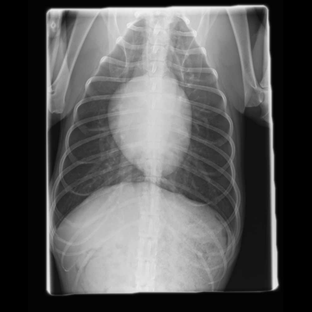
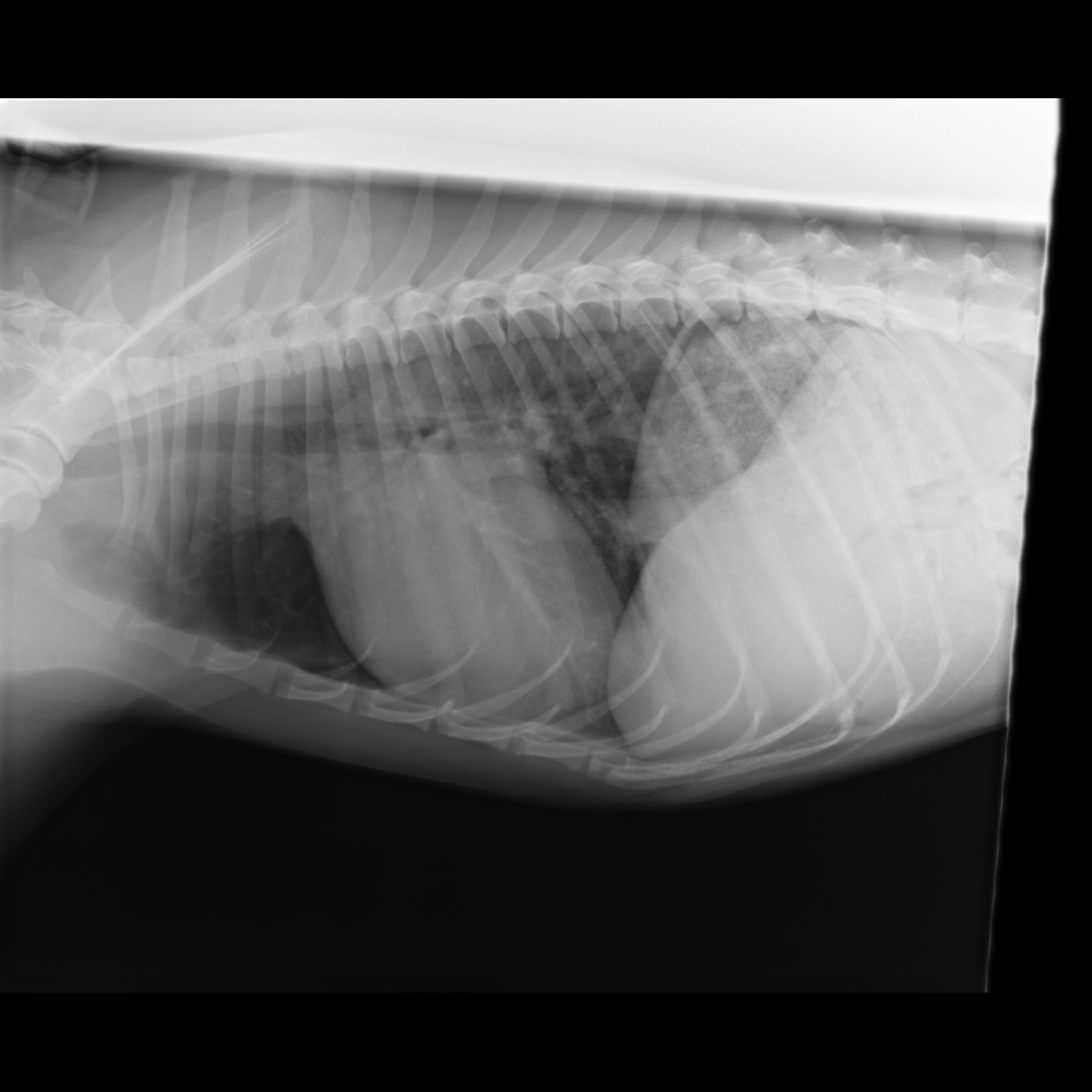
 Radiographs
Click here for ventral dorsal view
 Clinical Labs
SERUM CHEMISTRIES
BUN: 19 mg/dL, Normal: <30 mg/dLCreatinine: 1.1 mg/dL, Normal: <2.1 mg/dL
Sodium: 144 mEq/L, Normal:138 – 154 mEq/L
Potassium: 4.4 mEq/L, Normal: 3.6 – 5.2 mEq/L
Chloride: 113 mEq/L, Normal: 105 – 119 mEq/L
ALT: 40 IU/L, Normal: <75 IU/L
ALP: 56 IU/L, Normal: <100 IU/L
HEARTWORM
Heartworm: Test Results NegativeURINALYSIS
Urinalysis – USG: 1.024Urinalysis – Protein: Negative
Urinalysis – Biochemical: Normal
Urinalysis – Sediment Evaluation: Norm
 Echocardiography
Please review the results of Hailey’s echo
Subjective – lesions of valves, myocardium, pericardial space: No pericardial effusion; normal systolic function of the left ventricle, thickened mitral leaflets with posterior leaflet mitral valve prolapse.
LV chamber size and thickness: Normal thickness but high normal to mildly increased chamber diameter.
Left atrial size: Mild dilation
LVIDd & LVIDs: Diastole (3.4 cm); systole (2.2 cm).
LV shortening fraction: Normal – 35%
RA, RV and Pulmonary Artery: Not remarkable (detailed images not shown).
Effusions: None.
Doppler results: Mitral regurgitation (MR); eccentric jet of MR is typical of primary valve disease; velocity of MR predicts normal systemic pressures; No tricuspid valve regurgitation (TR). Normal mitral inflow velocities.
 ECG
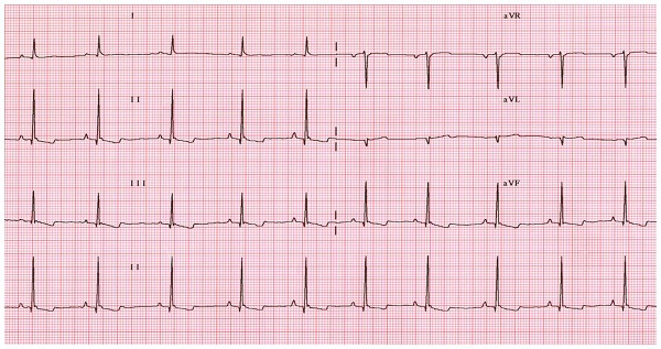
ECG
Technical Quality, Leads, Paper Speed, Calibrations: Satisfactory recording; 6 frontal leads and lead 2 rhythm strip recorded; paper speed 50 mm/sec; calibration 10mm/mV.
Artifacts: No significant
Rhythm- regular or irregular/ patterns: Regularly irregular. Heart rate 110 bpm.
Heart Rhythm Disturbances: Respiratory sinus arrhythmia; no premature complexes were detected.
P Wave Abnormalities- morphology, amplitude, duration: Normal P wave height and width.
QRS Abnormalities- axis, morphology, amplitude, duration: Normal frontal axis, no significant increase in height or width (insensitive measurement of chamber size).
Abnormal Intervals- PR, QRS, QT: Normal intervals.
Other: Slurred ST segments.


