Whitley
 Case Background
Age: 11 years
Sex: Male, neutered
Breed: Cocker Spaniel
 Clinical History
The patient had brief anesthesia two days ago for a dewclaw injury. He began coughing and hacking on day of surgery and has not improved since. On physical examination today:
- HR- 120 bpm.
- Normal pulse strength, no deficits.
- A grade 4/6 left apical systolic murmur was detected as well as a 3/6 right apical systolic murmur.
- Whitley had labored breathing at an increased rate, and pulmonary crackles were auscultated over all lung fields.
 Radiographs
Click here for Whitley’s radiograph viewer (Measure VHS and VLAS)
Technical details
Location of images: Thoracic radiographs obtained.
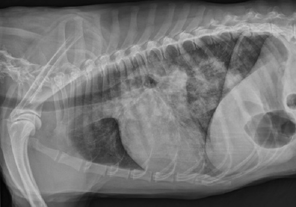
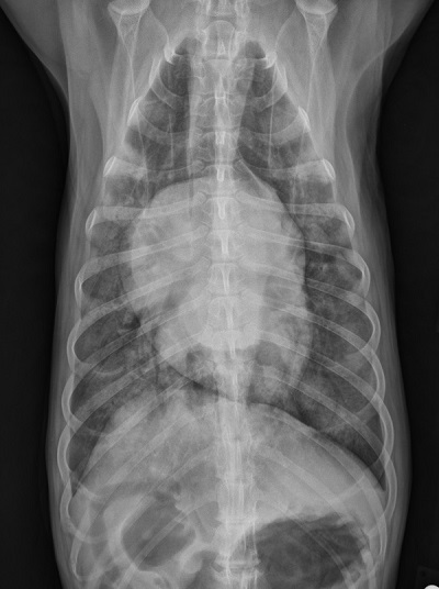
Views of images: Left lateral and ventrodorsal throracic radiographs were obtained.Radiographic findings
Technical issues: Good exposure and positioning.
Cardiac size including VHS: VHS=11; Moderate left ventricular and severe left atrial enlargement.
Other findings: Severe dorsal caudal and ventral caudal alveolar infiltrates; a milder alveolar pattern is visible in the cranial ventral right lung lobe on the lateral view. The pulmonary veins are enlarged.
 Diagnosis & Treatment
Discussion: The radiographic findings of severe alveolar infiltrates paired with left-sided heart enlargement and pulmonary venous enlargement are consistent with left-sided congestive heart failure with pulmonary edema. The combination of clinical findings and radiographic findings are suggestive of mitral valve insufficiency as the underlying etiology, although other causes of left heart failure cannot be ruled out based on radiographs alone. An echocardiogram was obtained, and mitral and tricuspid insufficiency due to chronic (myxomatous) valve disease was diagnosed.
Treatment/management: The patient was hospitalized in an oxygen enriched environment. Intravenous furosemide and oral pimobendan were administered and the patient was greatly. improved within 18 hours and released with oral furosemide, pimobendan and enalapril therapy. One week later, the patient was “back to normal” at home, and radiographs (“recheck”) were obtained. These radiographs (left lateral and VD view) show continued left ventricular and left atrial enlargement but complete resolution of the previous alveolar pattern (pulmonary edema).

