
Chloe
 Case Background
Name: Chloe
Age: 14 months
Sex: Female, intact
Breed: Golden retriever
Age: 14 months
Sex: Female, intact
Breed: Golden retriever
 Clinical History
Please review Chloe’s clinical history.
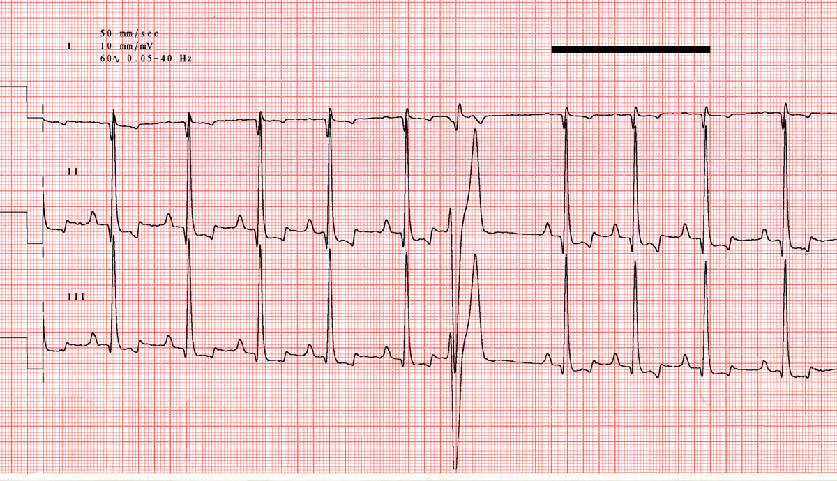
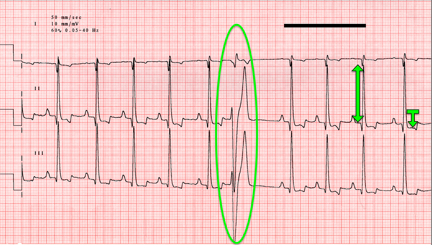
Chloe had an abnormal heart rhythm heard on exam. She also has a loud left basilar systolic murmur and weak femoral pulse quality.
 ECG
ECG
View Chloe’s electrocardiogram (the black bar represents one second)
What abnormalities are present on this ECG?
 Discussion & Treatment
Discussion: These changes suggest a disease process affecting the left ventricle. In a young golden retriever with a left basilar heart murmur and weak pulse quality, subaortic stenosis would be the top differential diagnosis, which would require echocardiographic evaluation for confirmation and to assess disease severity. Treatment/management: A single PVC may not require treatment; if signs of syncope are noted or longer ECG recordings (e.g., a 24-hour Holter monitor) show more malignant runs of ventricular ectopy, then antiarrhythmic therapy would be considered. If subaortic stenosis is confirmed on echocardiography with a moderate or severe gradient, atenolol therapy may be considered for cardioprotection and potential antiarrhythmic effects.