
Isabella
 Case Background
Name: Isabella
Age: 5 months
Sex: Female
Breed: Yorkie
Age: 5 months
Sex: Female
Breed: Yorkie
 Clinical History
Please review Isabella’s clinical history.
- Essentially normal puppy
- Loud systolic murmur
- Owner wants to have Isabella spayed
- Radiographs taken as part of anesthetic evaluation
 Radiographs
View Isabella's right lateral radiograph
 View Isabella's ventrodorsal radiograph
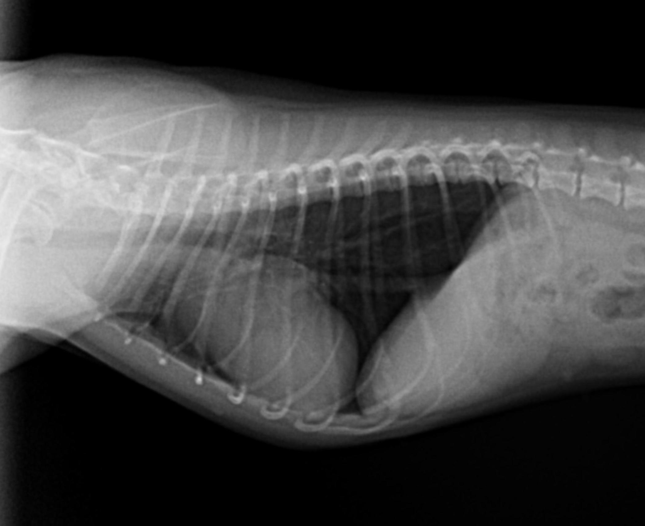
Isabella's right lateral thoracic view
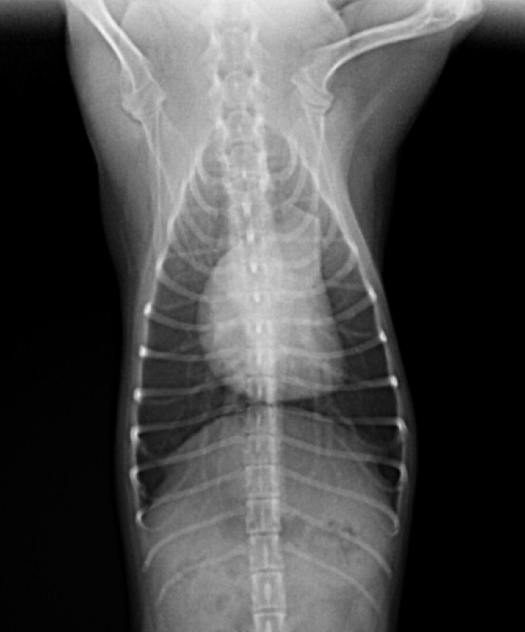
Isabella's ventrodorsal thoracic view
Technical details
Location of images: Thoracic radiographs obtained.
Views of images: Right lateral and ventrodorsal views.Radiographic findings
Technical issues: Well positioned radiograph, good technique, inspiratory film.
Cardiac size including VHS: VHS is at the upper limits of normal (10.6); subjectively the right side may also be enlarged (wide on lateral and VD suggests some RVE); The most striking abnormality on the film is the dilated pulmonary artery (at the 1:00-2:00 position on the VD).
Other findings: The vascular patterns are normal, but may be slightly reduced (a very subjective assessment). No abnormalities are associated with the pleural space or pulmonary parenchyma.
 Diagnosis & Treatment
Radiographic interpretations: Mild cardiac enlargement, involving mostly the right side; prominently enlarged main pulmonary artery (most apparent on the VD, suggestive on the lateral); normal to small left side; normal pulmonary parenchyma with normal to small pulmonary vessels.
Discussion: The presence of an enlarged right side coupled with a prominent main pulmonary artery is most consistent with pulmonic stenosis. Other abnormalities that are associated with a dilated main pulmonary artery would either be associated with a large left side (VSD, PDA) lack of a loud systolic murmur (ASD), or be associated with pulmonary hypertension and more significant clinical signs (right to left shunting of blood flow, heartworm disease).
Treatment/management: Therapy at this stage would be premature; an echocardiogram would ideally be performed to confirm the disease and estimate its severity. Based on these radiographs, there is no overt contraindication for anesthesia, but the anesthetic protocol may be altered depending on the echo findings/final diagnosis.