
Kodiak
 Case Background
Name: Kodiak
Age: 7 year old
Sex: Female, spayed
Breed: Siberian Husky
Age: 7 year old
Sex: Female, spayed
Breed: Siberian Husky
 Clinical History
Please review Kodiak’s clinical history.
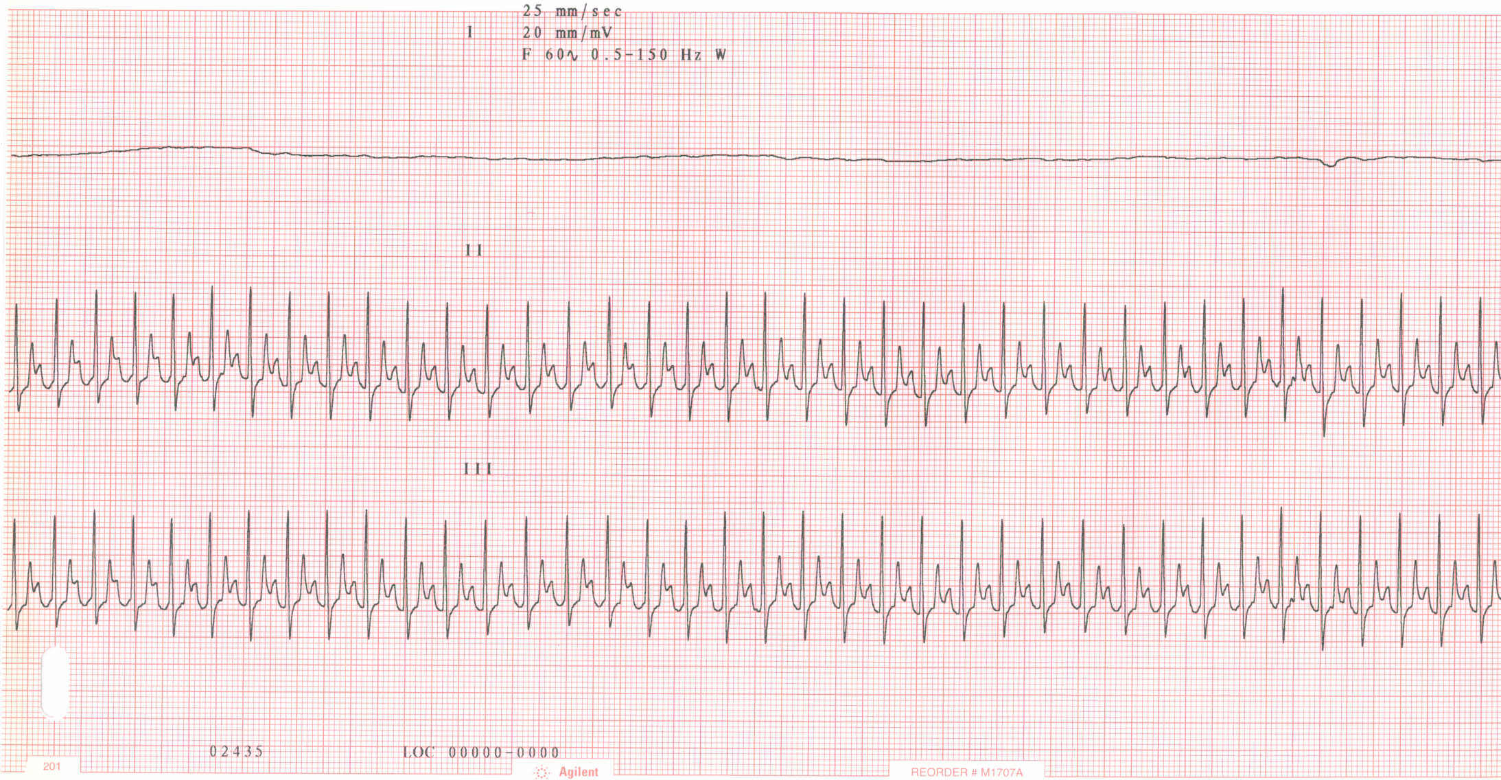
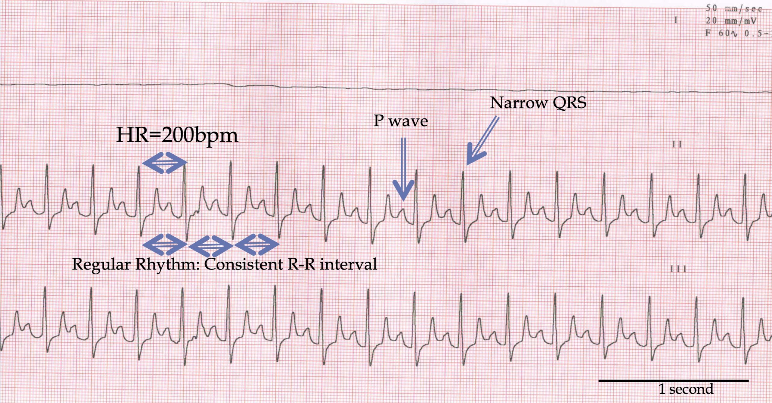
Patient presented 30 hours after an ovariohysterectomy. Patient is recumbent with pale MM, weak femoral pulse, a soft heart murmur, and fluid in the abdomen detected via ultrasound examination. Blood pressure was 80mmHg systolic.
 ECG
ECG
View Kodiak’s electrocardiogram (the black bar represents one second)
What abnormalities are present on this ECG?
 Discussion & Treatment
Discussion: Sinus tachycardia is the logical diagnosis, given the recent history of abdominal surgery and fluid in the abdomen. The most likely cause of the sinus tachycardia is systemic hypotension secondary to arterial bleeing into the abdomen (hypovolemic shock). The natural response to hypotension is an increase in sympathetic tone which increases the sinus rate. Treatment/management: Administer shock dose of fluids, perform a cystocentesis to confirm the fluid in the abdomen is blood and perform emergency abdominal exploratory surgery, looking for a bleeding uterine or ovarian artery.