AJ
German Shepherd
Select Radiograph(s)

Radiographic Report
Visit 1:
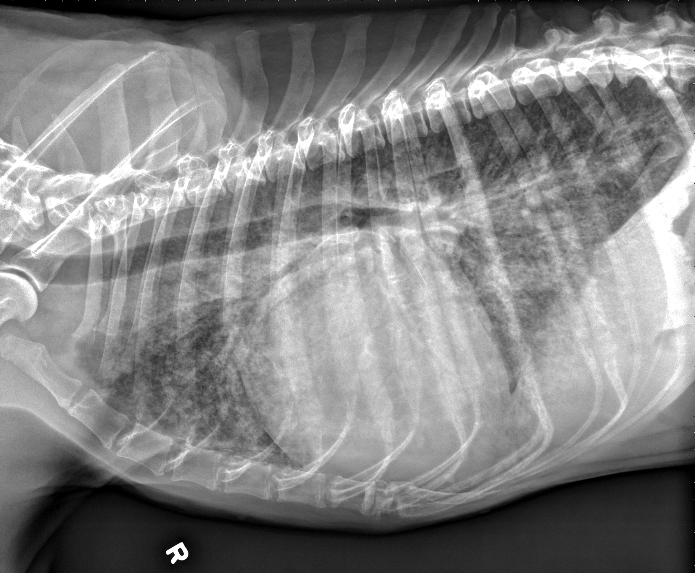
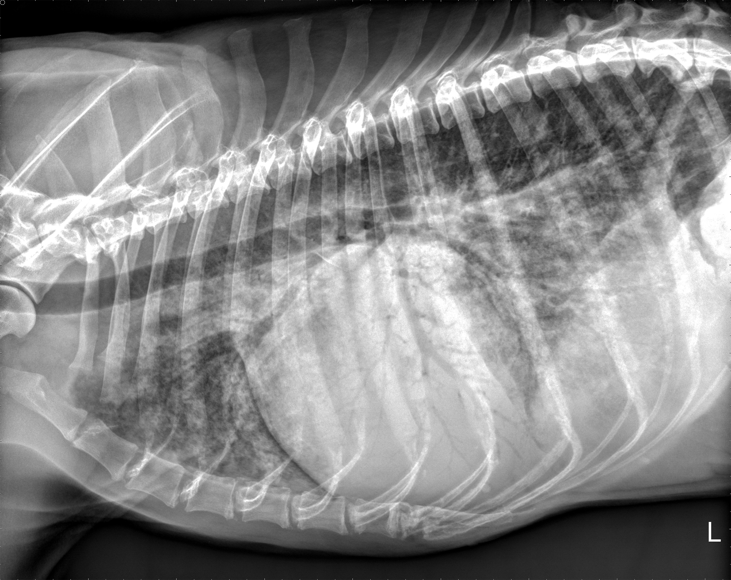
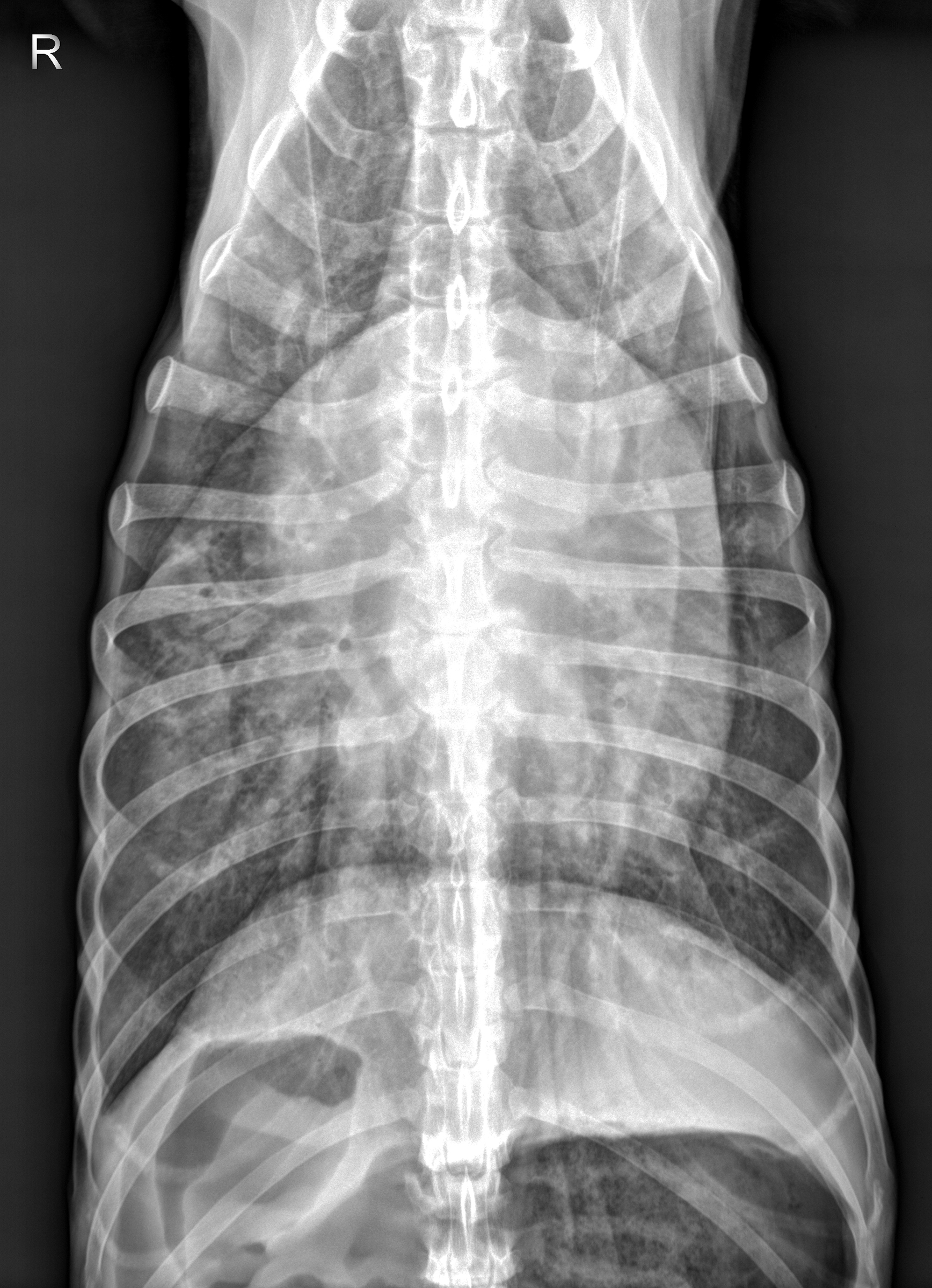
Radiographic interpretation:
There is an interstitial to alveolar pattern in all lung lobes, more severe in the right middle lung lobe creating air bronchograms and lobar sign with the adjacent lobes. The interstitial pattern is predominately peribronchial in distribution. There is also moderate to marked enlargement of the cranial lobar veins, while the caudal pulmonary vessels are poorly defined. There is moderate generalized cardiac enlargement, occupying 4 intercostal spaces on the lateral images and greater than 2/3rd the thorax on the VD image. There is a mild caudodorsal cardiac bulge with round double opacity in the region of the left atrium on the VD. The pleural space, mediastinum, and diaphragm are within normal limits. The included musculoskeletal structures are normal.
Conclusion: Diffuse peribronchial interstitial to alveolar pattern and generalized cardiomegaly with mild left atrial enlargement most likely represents left-sided congestive heart failure.
Clinical interpretation/ additional case information:
This dog presents with signs, physical findings, and radiographic changes consistent with left-sided congestive heart failure. AJ received intravenous butorphanol for sedation and an intravenous dose of furosemide and was placed in an oxygen cage with 40% inspired oxygen. Once his respiration rate and effort improved, a noninvasive blood pressure measurement was taken; his systolic blood pressure measured by Doppler methods was 112 mmHg (systolic blood pressure by Doppler). A brief echocardiogram was performed four hours after admission and demonstrated severe mitral valve regurgitation, enlarged left atrium and left ventricle, and markedly reduced left ventricular systolic function (fractional shortening = 15%, normal> 25%). AJ was treated for 24 hours in-hospital with intravenous furosemide, oral pimobendan, and supplemental oxygen with gradual improvement in his respiratory signs. Thoracic radiographs were repeated 10 hours later (visit #2).
Visit 2:
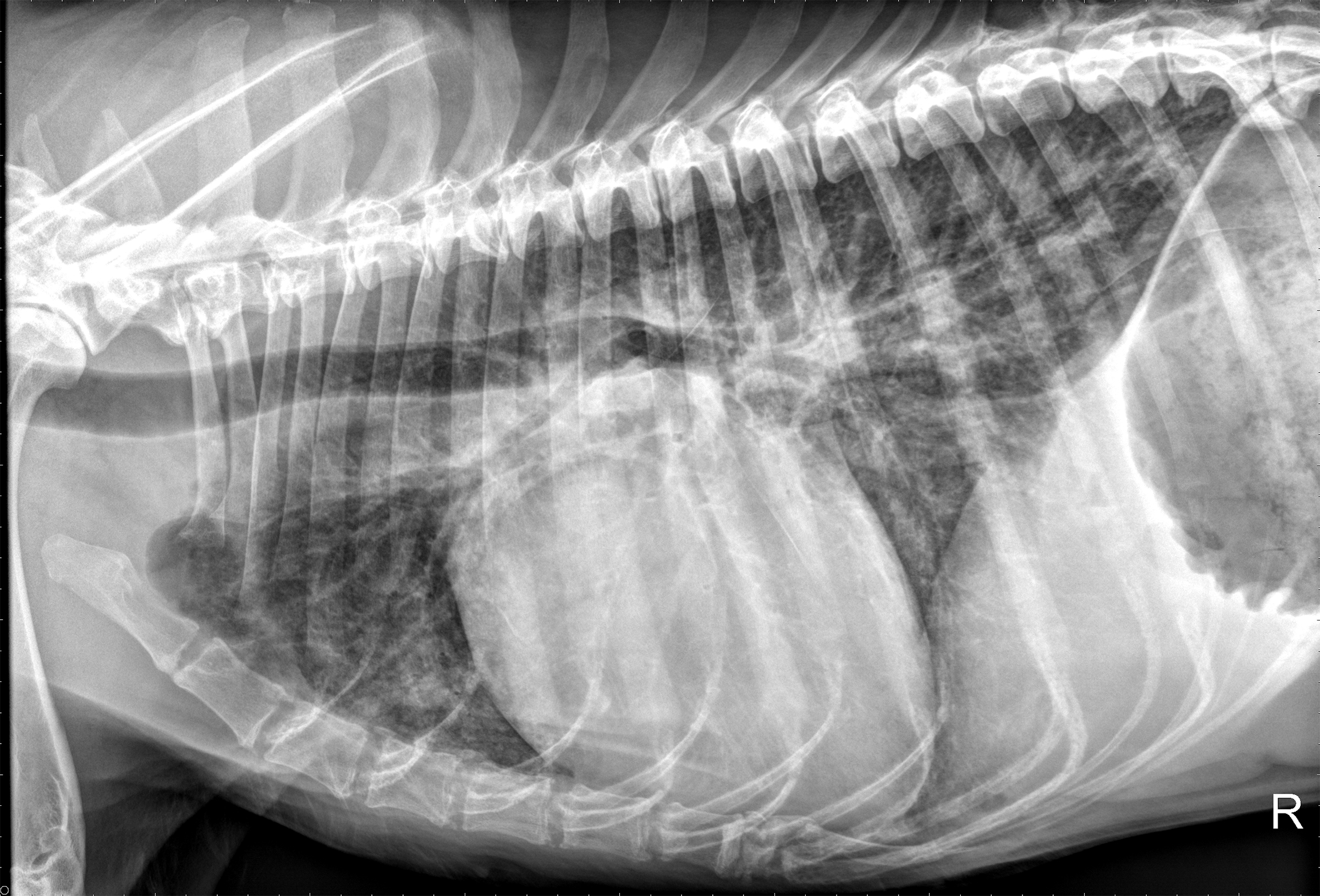
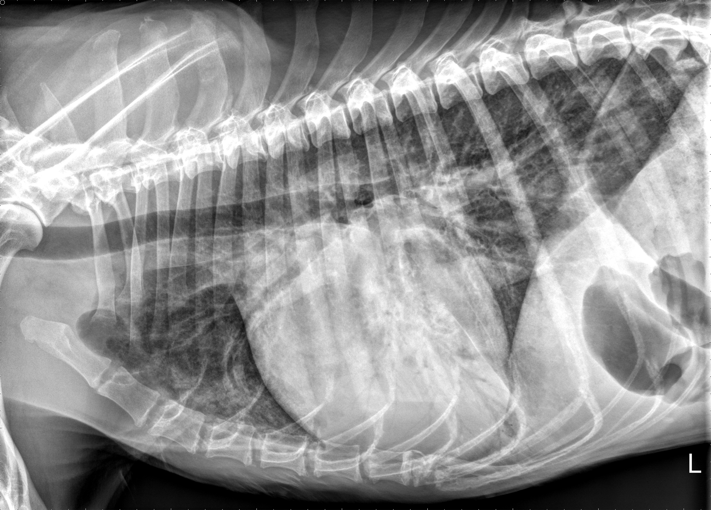
Visit #2 Radiographic interpretation:
There is an improved peribronchial interstitial to alveolar pattern in all lung lobes. The interstitial pattern remains. The dorsal alveolar pattern in the right middle lung lobe has improved, with persistent ventral alveolar pattern. There is improved pulmonary venous distension (less dilated). There is similar cardiomegaly and left atrial enlargement, though overall VHS has decreased slightly (likely related to diuresis or improved detection of cardiac borders). The pleural space, mediastinum, and diaphragm are normal. There is moderate gas in the stomach. The remainder of the exam is unchanged.
Radiographic Diagnosis: Improved interstitial to alveolar pattern affecting all lung lobes likely representing therapeutic response of cardiogenic pulmonary edema to furosemide/pimobendan therapy. The residual pulmonary infiltrates likely represent persistent cardiogenic pulmonary edema.
Clinical interpretation/ additional case information:
This dog was presented with signs, physical findings, and radiographic changes consistent with left-sided congestive heart failure. Serial bloodwork was largely unremarkable, though hyponatremia, hypokalemia, and hypochloremia developed throughout the hospitalization. Home therapy included furosemide and pimobendan; enalapril and spironolactone were started 2 weeks later once appetite had normalized.
Clinical History
Signalment: 11-year-old MC German Shepherd Dog
Clinical History
AJ is an 11-year-old German Shepherd who was presented to an emergency practice for evaluation of respiratory distress and weight loss. He had a cough that developed 2 months ago and lasted two to four weeks before subsiding. In the past week, the coughing has returned and he has also developed a more labored breathing pattern. His breathing has gotten worse in the past 3 days and he has become more lethargic. He cannot lay down comfortably. The owners also reported that AJ had vomited bile last night. After he vomited, he appeared incoherent and wasn’t fully responsive, but snapped out of it after about 10 minutes.
On physical examination, he was thin with a body condition score of 2 out of 9. He was panting with increased effort, heart rate was 140 beats per minute. A grade III/VI left apical systolic heart murmur was auscultated and lung sounds were harsh with increased effort.
Visit 2:
Clinical History
These films are from the same 11-year-old German Shepherd as visit #1 and were taken 10 hours after starting therapy (oxygen, furosemide, pimobendan).