Albert
Miniature Schnauzer
Select Radiograph(s)


Radiographic Report
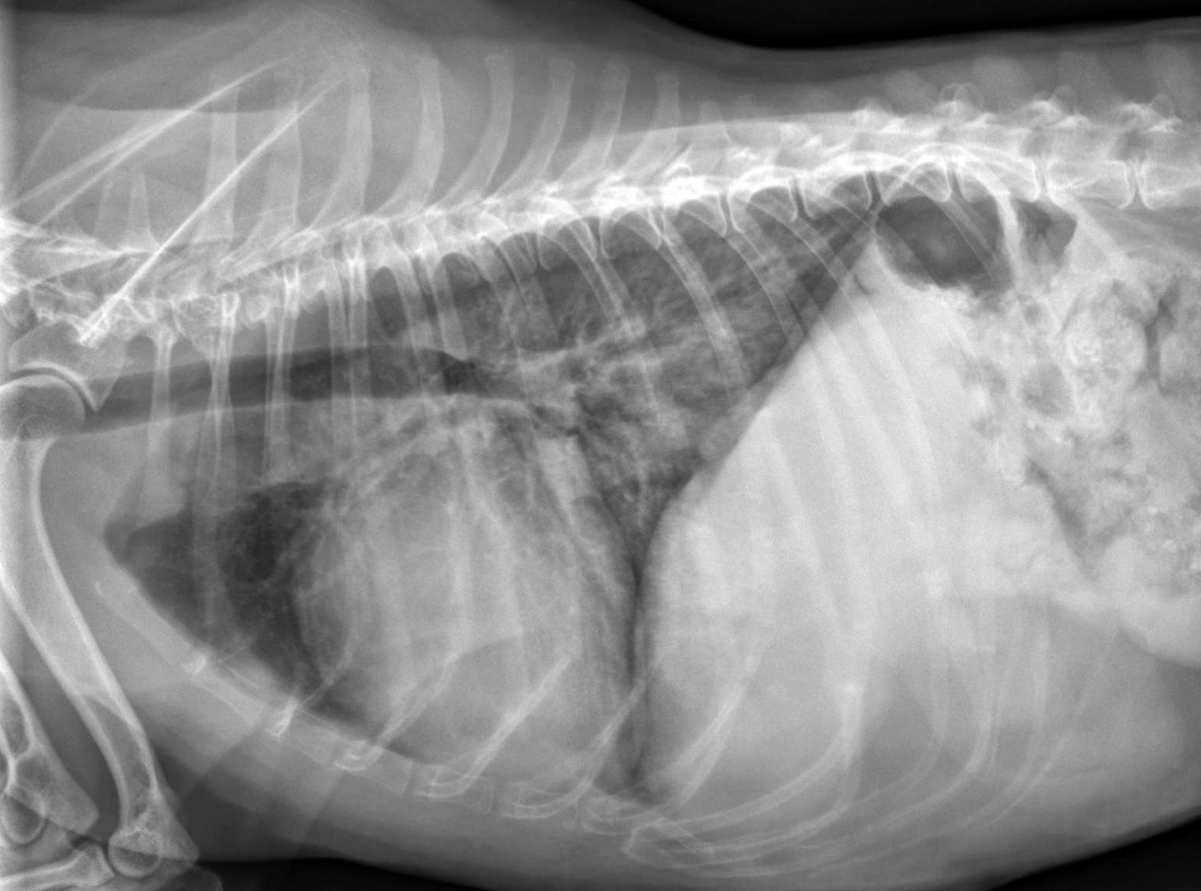
Radiographic interpretation: There is cardiomegaly characterized mainly by elongation of the heart. The VHS is either at the upper limit of normal for the breed (no breed normal published) or is mildly increased. Subjectively the left atrium is mildly enlarged but the VLAS is only at the upper limits of normal. The main pulmonary finding is an increased intrapulmonary density, with air bronchograms, in the caudal lung lobes. These alveolar infiltrates are best seen on the VD projection (see the annotated VD image, arrows). Although this dog had clinical evidence of valvular heart disease, the respiratory distress was thought to be related to noncardiogenic pulmonary edema associated with a brain tumor and seizure disorder. The distal distribution of infiltrates, more severe away from the hilus of the heart, supports this suggestion. Typical of the left lateral view, the cardiac apex is more elevated and both the left atrium and cranial pulmonary vein are more prominent; these are not seen on the right lateral projection.
Clinical interpretation / additional case information This patient’s clinical diagnosi is challenging because of the preexisting mitral valve disease, a heart murmur and associated cardiomegaly, along with an acute onset of respiratory distress that is similar to that seen with mitral chord ruptures. However, the association of noncardiogenic (neurogenic) lung edema after seizures in dogs is well known and this type of edema is typically distributed in the periphery of the caudo-dorsal lobes. Possible mechanisms are redistribution of blood flow to the lungs and increased pulmonary capillary permeability. This 13-year old dog was euthanatized after initial evaluation due to progressive neurologic deterioration.
Clinical History
Signalment: 13 year old MN Miniature Schnauzer
Clinical History Presented for respiratory distress after multiple seizures earlier in the day. Patient has a history of chronic compensated mitral valve disease and is not currently receiving any cardiac medications. Albert has experienced progressive behavioral changes and vocalization over the past month. Examination indicates loud bronchial sounds bilaterally, increased respiratory effort, and a grade 3/6 systolic murmur loudest over the left apex. Neurological consultation suggested a frontal lobe lesion, probably a neoplasm, to explain the seizures and behavioral changes. Initial clinical management included midazolam, oxygen, and one injection of furosemide. Radiographs were obtained.