Brownie
Mix Breed
Radiographic Report
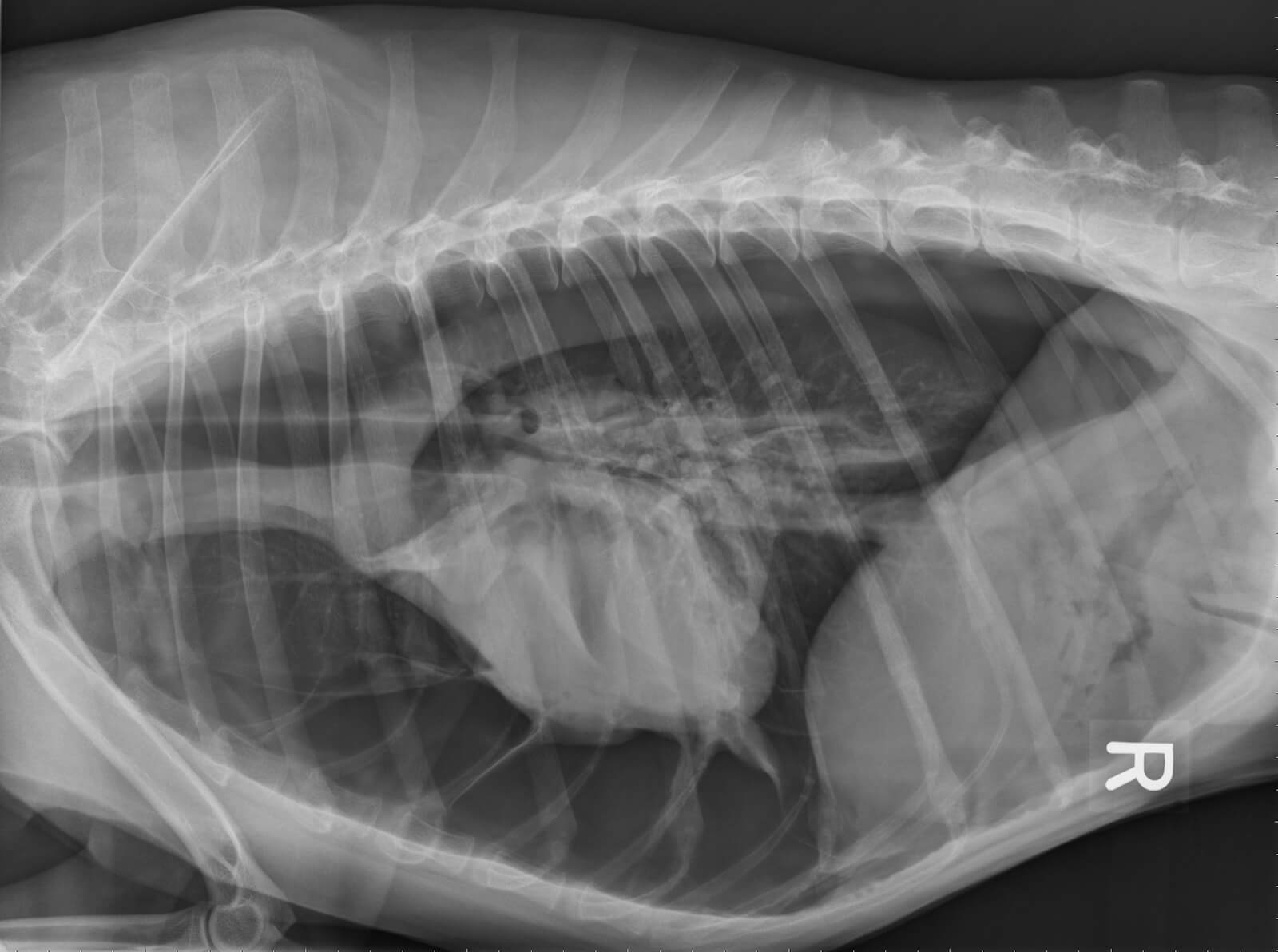
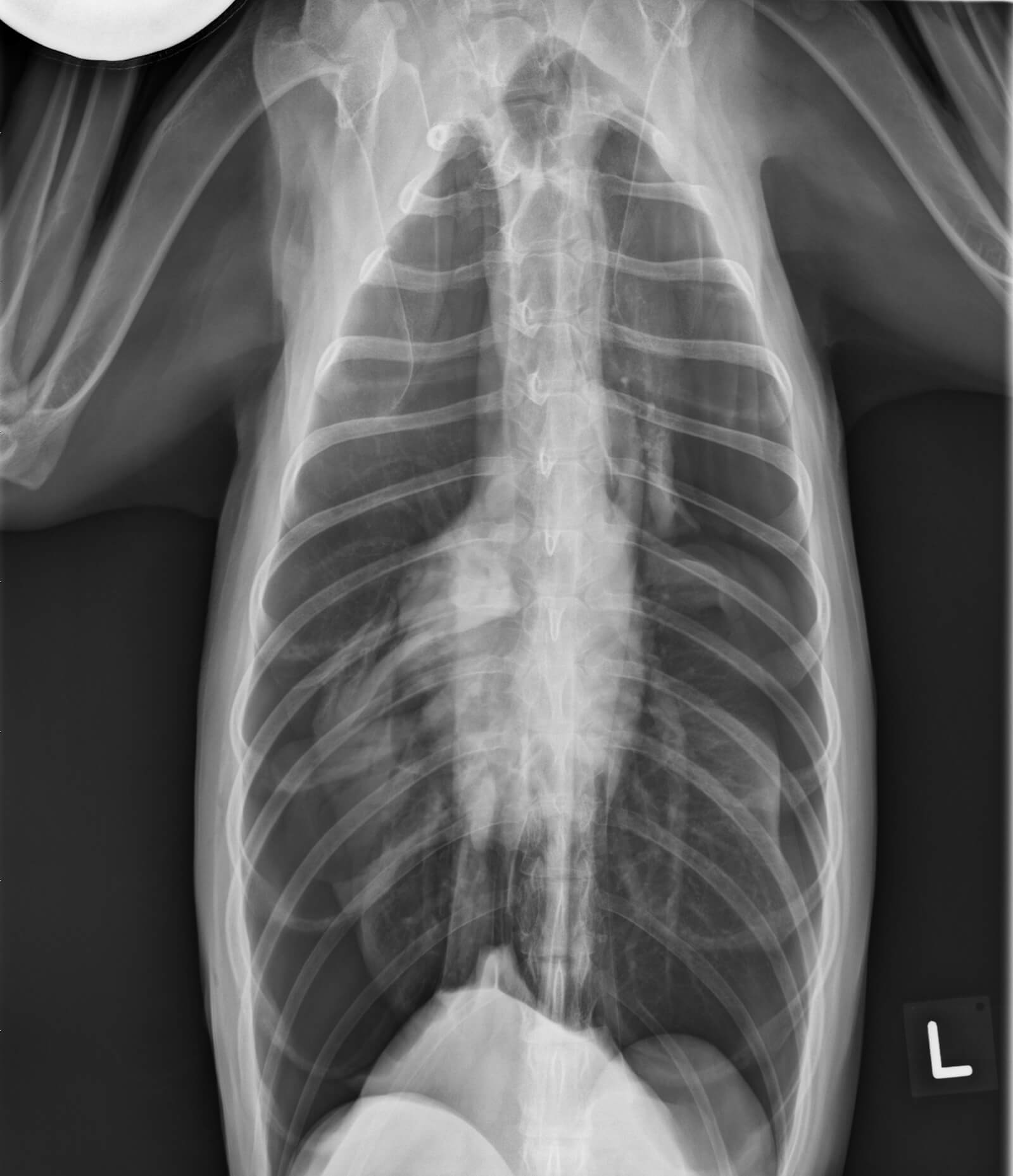
Radiographic interpretation: Cardiac size is greatly reduced and the cardiac silhouette is retracted away from the sternum (yellow arrows on annotated image). The VHS cannot be calculated because the edges of the cardiac silhouette are obscured by overlying lung and mediastinal tissue. Free gas is visible in the thorax extending to the diaphragm, and the retracted edges of the underinflated lung lobes are visible (red arrows on annotated image). There is no evidence of thoracic trauma (broken ribs, soft tissue swelling). These findings are consistent with a pneumothorax.
Additional information: Immediate thoracocentesis is required to remove air from the thoracic cavity. Chest tube placement and continuous suction may be necessary to acutely stabilize a pneumothorax.
Clinical History
Signalment: 4 year old MC Mixed breed dog
Clinical history Presented on emergency service for acute onset difficulty breathing. No previous medical problems. No history of trauma. Heart rate on presentation 180 bpm and regular. Pink mucous membranes, tachypneic