Coconut
Domestic Shorthair
Select Radiograph(s)
Radiographic Report
Radiographic interpretation:
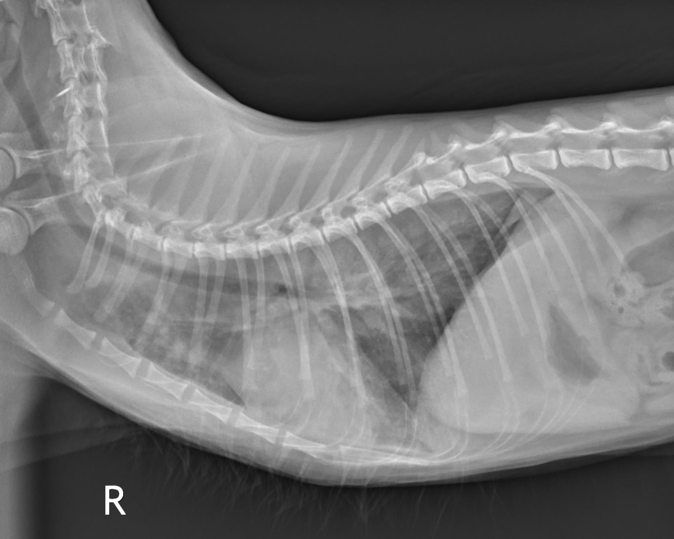
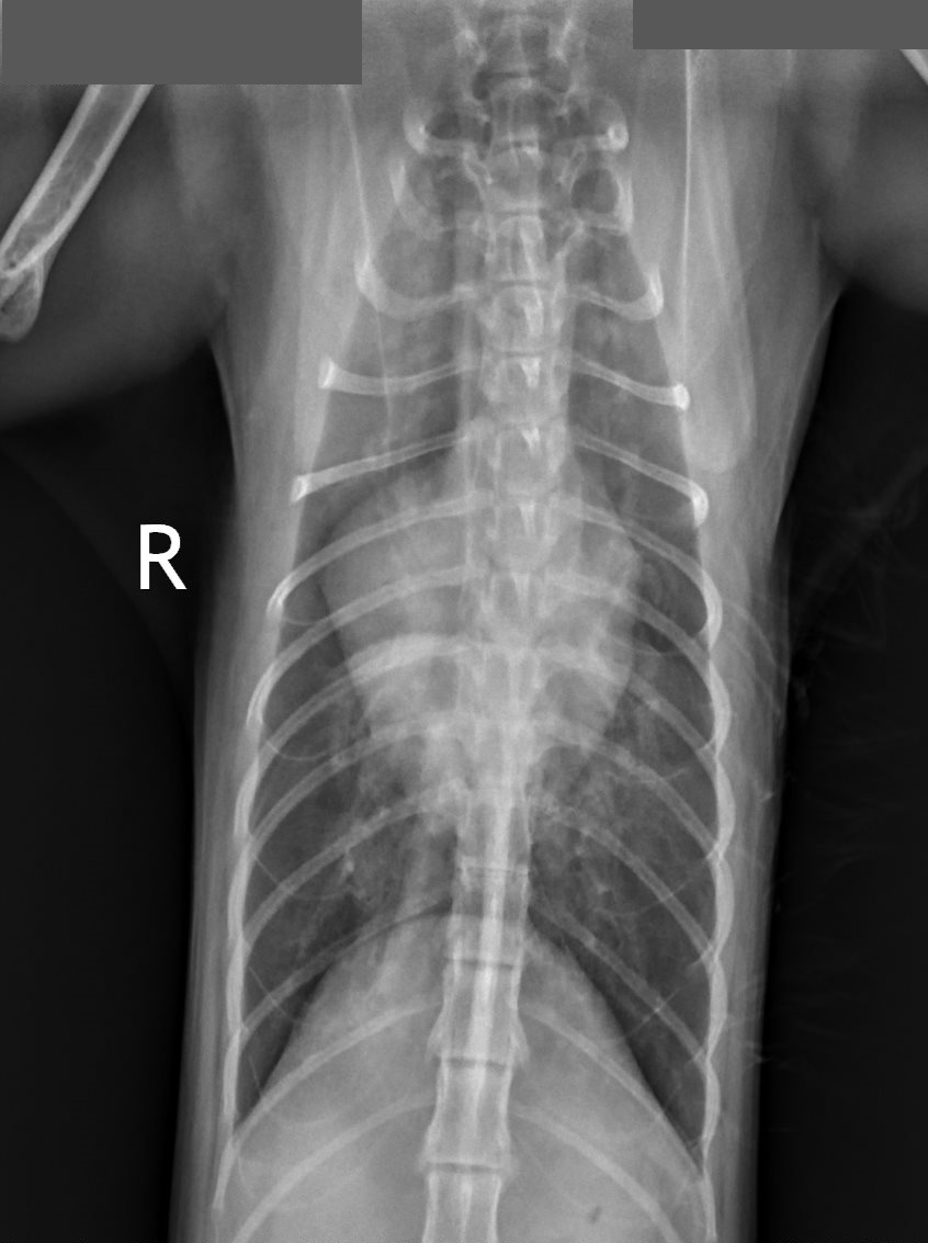
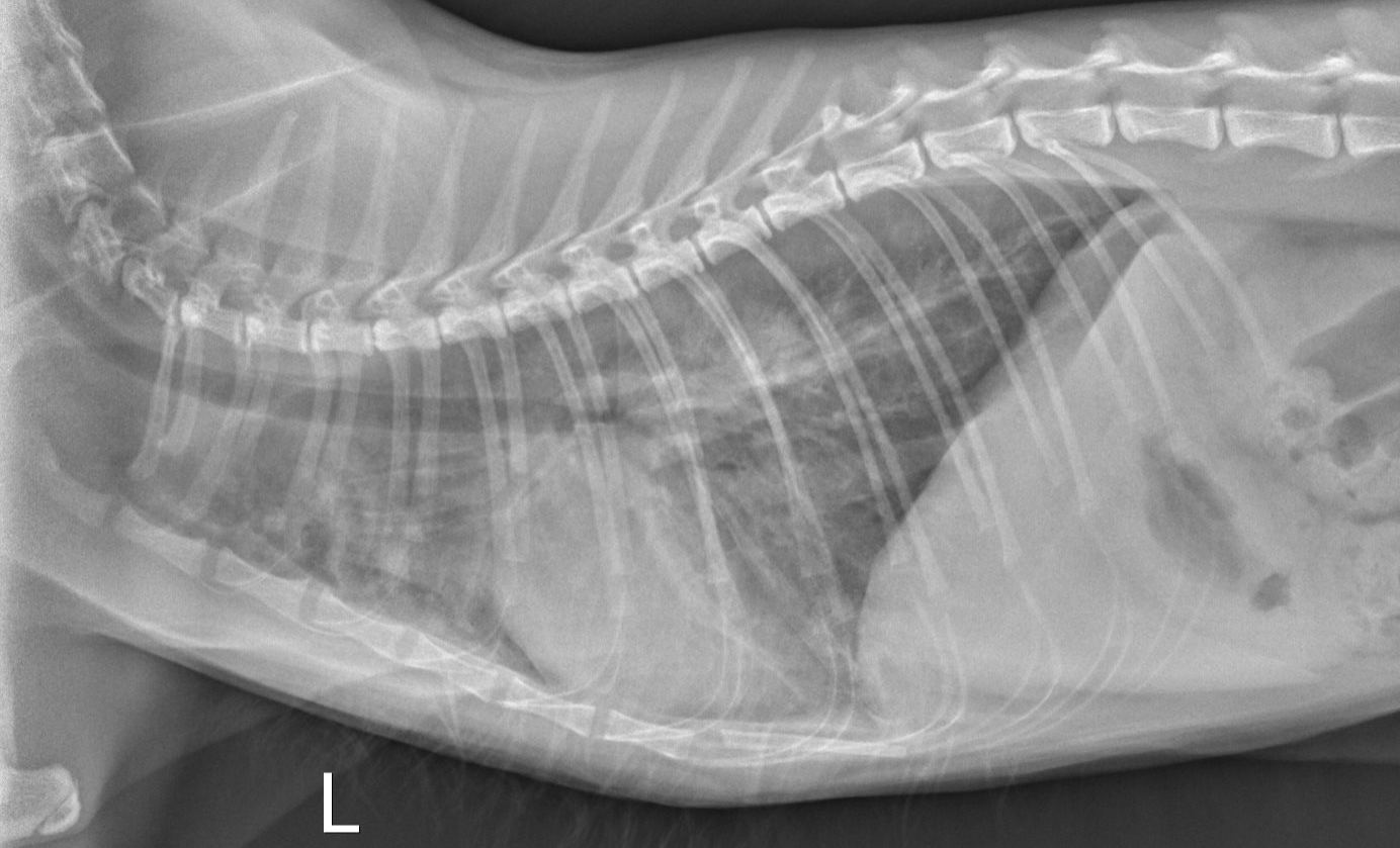
Severe cardiomegaly with obvious atrial dilation is noted on the VD view. There is significant pulmonary venous distention and loss of vascular detail. There is a heavy perihilar/caudodorsal interstitial pattern bilaterally. Findings consistent with congestive heart failure.
Clinical interpretation / additional case information:
An echocardiogram revealed findings consistent with hypertrophic cardiomyopathy. There was spontaneous echo contrast (“smoke”) identified in the markedly dilated left atrium. The patient was treated acutely with IM furosemide and oral pimobendan, and showed marked clinical improvement by Day 2. Oral enalapril and clopidogrel were added to therapy for discharge.
Clinical History
Coconut is a 10yr old, male neutered, Domestic Shorthair with no previous history of cardiac disease. He recently had a normal Total T4 at 2.4mg/dl. The family arrived home after work and noticed the patient was tachypneic with increased respiratory effort. The family rushed him to the veterinary emergency hospital. Upon presentation, a gallop heart sound was detected, but no heart murmur was identified. The Doppler systolic blood pressure was high normal at 150mmHg . There were also bilateral pulmonary crackles noted.