Cornelius
DSH
Select Radiograph(s)
Radiographic Report
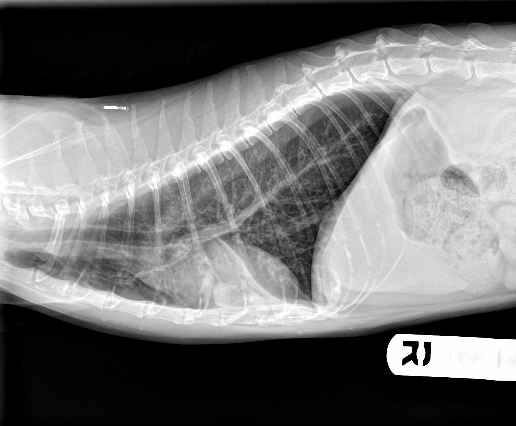
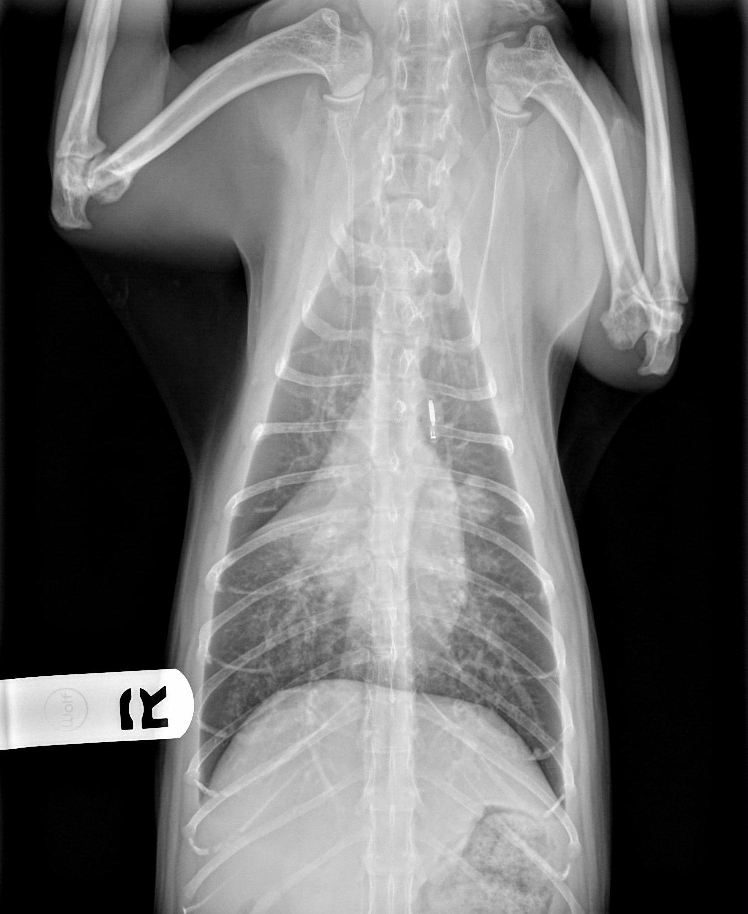
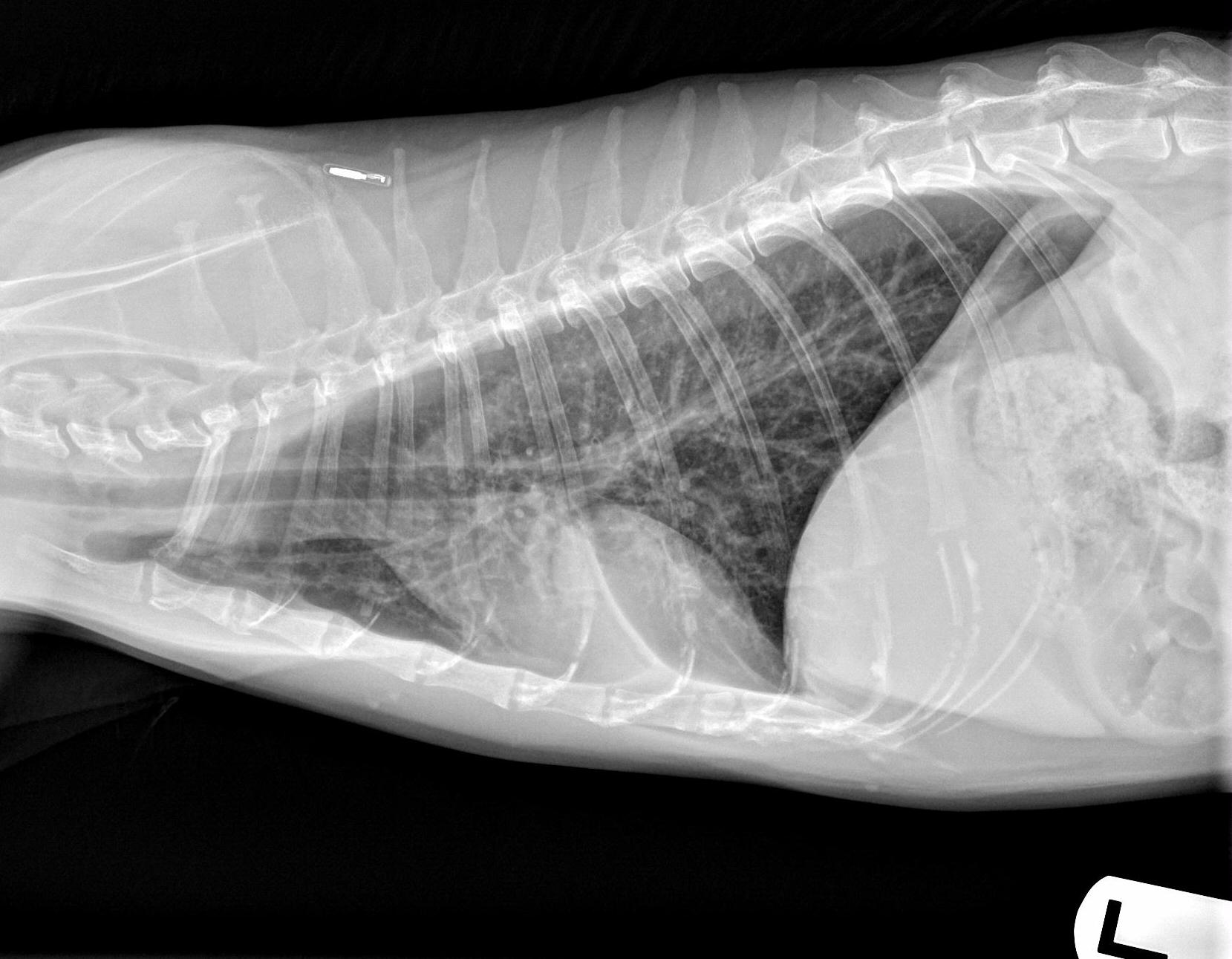
Views of images: Left and right lateral and ventrodorsal throracic radiographs.
Radiographic Findings
Technical issues: Good technique.
Cardiac size including VHS: VHS=7.9; Cardiac silhouette appears subjectively normal for an older cat in which the aorta elongates, changing the angle in which it enters the left ventricle and the cardiac silhouette increases its sternal contact. There is a bulge at the 12 o’clock position that is due to the same age related change in which the aorta elongates and the cardiac silhouette increases its sternal contact, making the aorta more visible cranial to the heart on a ventrodorsal radiograph. These patients have less overlap of the aortic arch and the cardiac silhouette, resulting in the bulge, immediately cranial to the heart on the VD. This should not be mistaken for a mediastinal or heart base tumor.
Other Findings: No pulmonary venous congestion, but the right main pulmonary artery appears to be slightly larger than the left (VD). Diffuse bronchial pattern, Alveolar Pattern with a “lobar sign” in the right middle lung lobe.
Radiographic Interpretation: Pneumonia of the right middle lung lobe. Suspected chronic feline asthma. Probable pulmonary hypertension.
Discussion: These films are most consistent with feline asthma and secondary lobar pneumonia with suspected pulmonary hypertension. The history of long standing asthma fits with the heavy bronchial pattern and predisposes this patient to recurrent pneumonia. Suspicion of pulmonary hypertension would also be high in a patient with chronic respiratory symptoms and current pneumonia. An echocardiogram would be needed to confirm pulmonary hypertension. A bronchoscopy revealed a mucus filled bronchus leading to the right middle lung lobe. Culture of this mucus identified Chlamydophila felisi that was susceptible to azithromycin.
Treatment/Management: For the pneumonia, the patient was placed on 6 days of 15mg of Azithromycin once daily with 20mg given on the first day, and Albuterol liquid 0.25mg every 8-12 hours. After one week, the patient was placed on a 3 week tapering dose of prednisonlone that started with 2.5mg every 12 hours for 7 days, then tapered down every 7 days until gone. This patient may require intermittent or sometimes chronic steroid and bronchodilator therapy. Antibiotics may be periodically needed given the expectation of chronic pulmonary compromise.
Clinical History
Name: Cornelius
Age: 13 years
Sex: Male, castrated
Breed: Feline, domestic short hair
Patient has a long standing history of cough and wheezing that has been treated with hydroxyzine periodically as well as prednisolone injections. However, the cough has been particularly bad lately and the character of the cough, now frequent, sounds wet and productive. He is also not eating well and has lost 1lb.


