Dana
Poodle
Radiographic Report
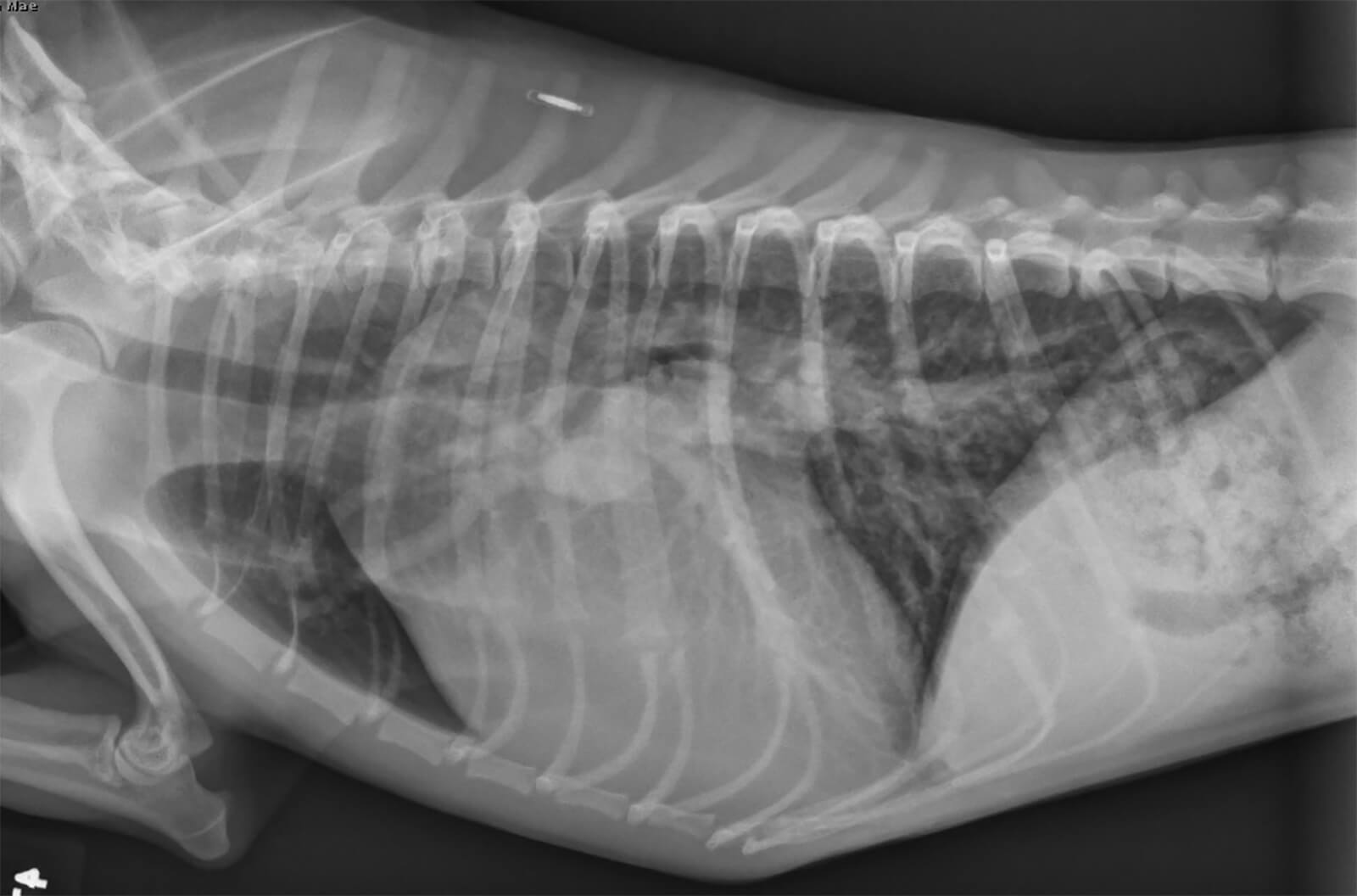
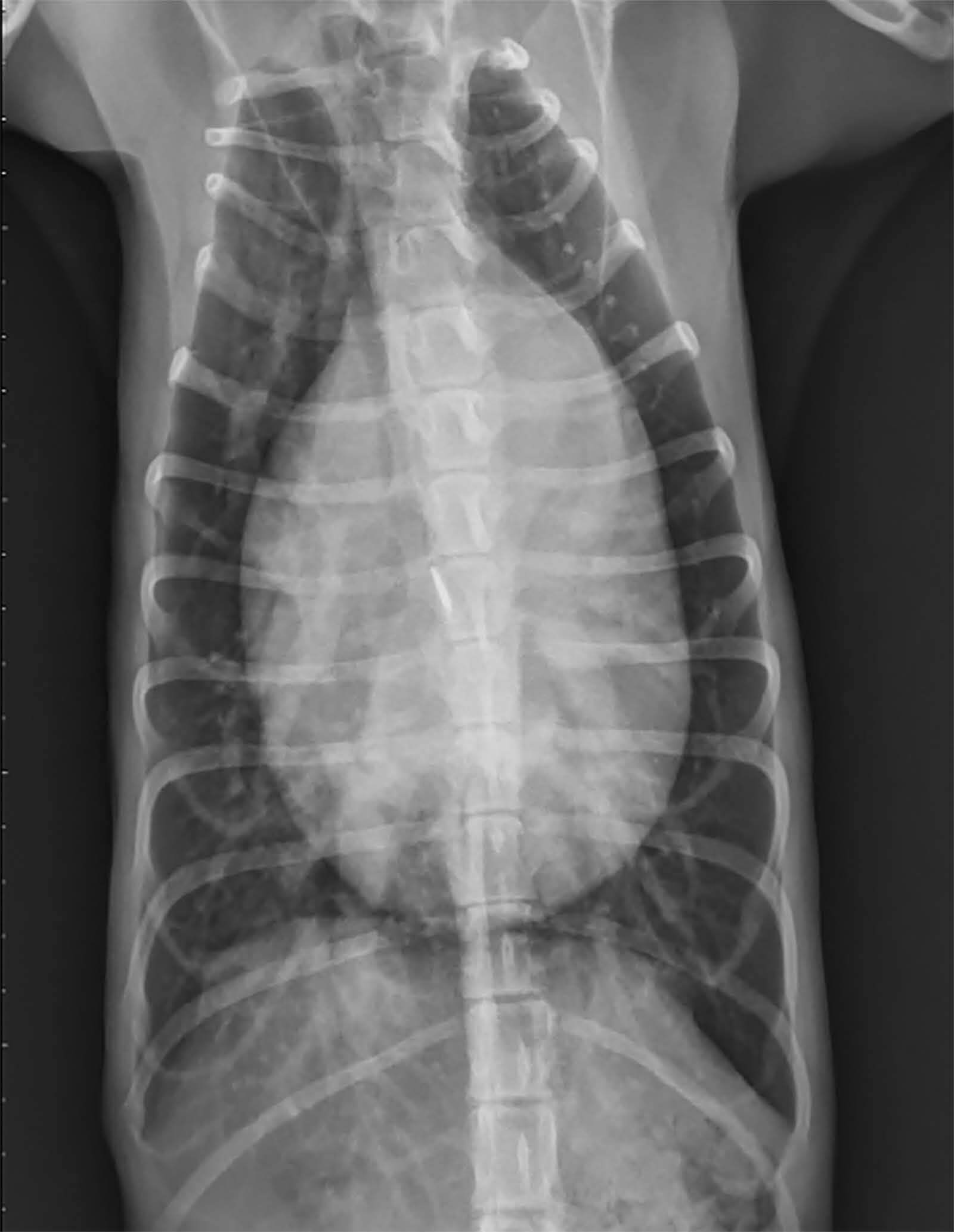
Radiographic interpretation: Severe enlargement of the cardiac silhouette characterized by left atrial and left ventricular enlargement. The main pulmonary artery is prominent on the lateral view coursing caudally from the heart base dorsal to the trachea. There is enlargement of pulmonary veins and pulmonary arteries (overcirculation pattern), consistent with a left-to-right cardiac shunt. The pulmonary fields show increased opacity due to the overcirculation pattern but no evidence of congestive heart failure is present.
Radiographic diagnosis: Findings are consistent with a patent ductus arteriosus (PDA), with pulmonary overcirculation but no congestive heart failure. Amplatz device placement via cardiac catheterization or surgical correction was recommended if echocardiographic examination confirmed the diagnosis.
Clinical History
Signalment: 2-3 year old F toy poodle mix
Clinical history: Presented for evaluation of a heart murmur. She was rescued from the streets 4 months ago and taken in by a rescue organization. Dana has been with her current foster family for the last 2 weeks. Her activity level is normal. She has not experienced any coughing, collapse, or exercise intolerance. She is eating and drinking normally. Dana is breathing comfortably at rest and has not had any episodes of respiratory distress. She is significantly underweight (BCS 3/9). A grade 5/6 continuous murmur was detected at the left heart base, with a 4/6 systolic murmur audible at the left heart apex. Her heart rate is normal (110 bpm) with bounding femoral pulses.