Duchess
Coonhound
Radiographic Report
Radiographic interpretation:
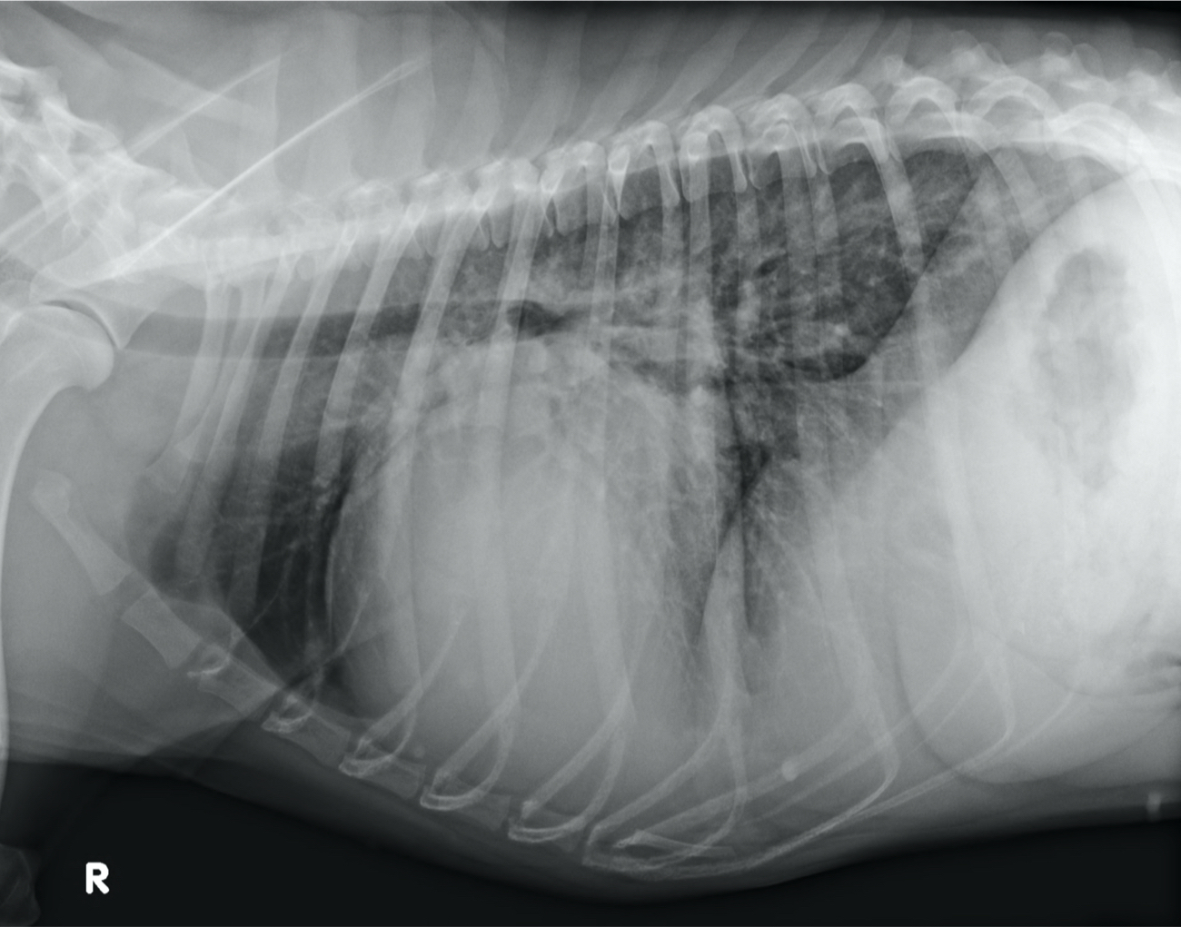
Lateral radiograph: Cardiomegaly is present with a VHS of 13.9. The radiograph suggests left ventricular enlargement with dorsal deviation of the carina and left atrial enlargement with loss of the caudal cardiac waist on the lateral view. The cranial cardiac silhouette is unremarkable and even though there is increased sternal contact the cardiac apex rests normally on the sternum. Therefore, although right ventricular enlargement often produces increased sternal contact, in this case the alteration may solely be related to the severity of left ventricular enlargement. There is a caudodorsal interstitial pattern present.
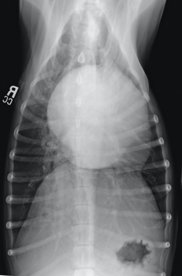
Ventrodorsal radiograph: The radiograph suggests left ventricular enlargement with prominence of the left ventricular apex from the 3 to 5 o’clock position. Both the left atrium (6 o’clock position) and the left auricle (3 o’clock position) appear enlarged. The superimposition of the enlarged left atrium and auricle over the remainder of the cardiac silhouette provides an increased opacity and the left atrial enlargement produces widening of the angle between the mainstem bronchi. The right atrium also appears enlarged in the 9 to 10 o’clock position. The caudal lobar pulmonary vasculature is distended with a faint interstitial pattern.
Radiographic diagnosis: Severe left ventricular and left atrial enlargement, right atrial enlargement, possible right ventricular enlargement, and pulmonary pattern suggestive of left-sided congestive heart failure.
Additional information: Duchess’ echocardiogram, ECG and radiographs were supportive of congestive heart failure secondary to atrioventricular myopathy and persistent atrial standstill. The owners elected to move forward with treatment despite the guarded long-term prognosis. A transvenous pacemaker was placed and Duchess was started on pimobendan, enalapril and furosemide. Although the disease was progressive she lived for another 3 years post-pacemaker implantation.
Clinical History
Signalment: 4 year old FS Coonhound
Clinical History: Duchess has lived with her owner since 12 weeks of age. She has an unremarkable past medical history and was last evaluated 8 months ago for routine preventative evaluation. At that time her cardiovascular physical examination findings were unremarkable. Duchess is now presented on emergency with a four-day history of coughing, lethargy and reduced appetite. Physical examination revealed a grade II/VI left sided systolic murmur, a heart rate of 42 beats per minute, strong femoral pulses, and mildly increased inspiratory lung sounds.