Duke
Ragdoll
Select Radiograph(s)
Radiographic Report
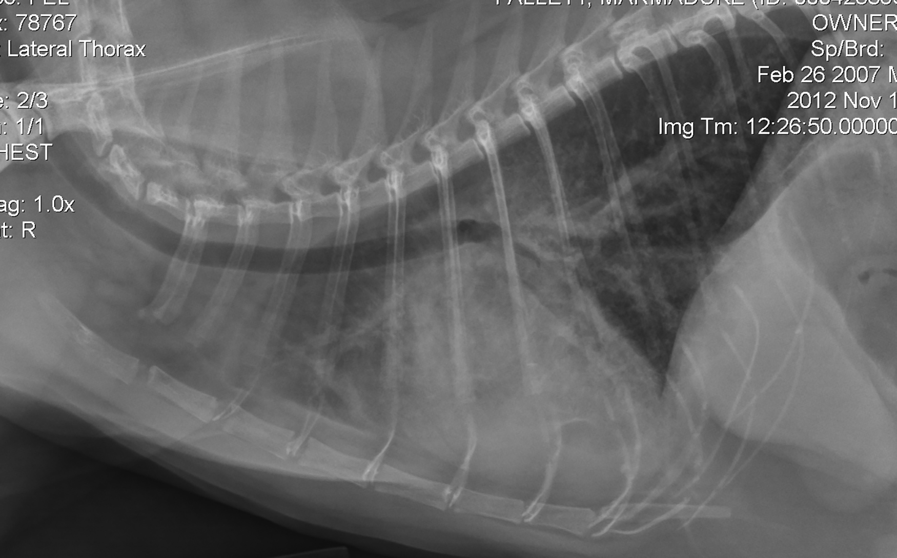
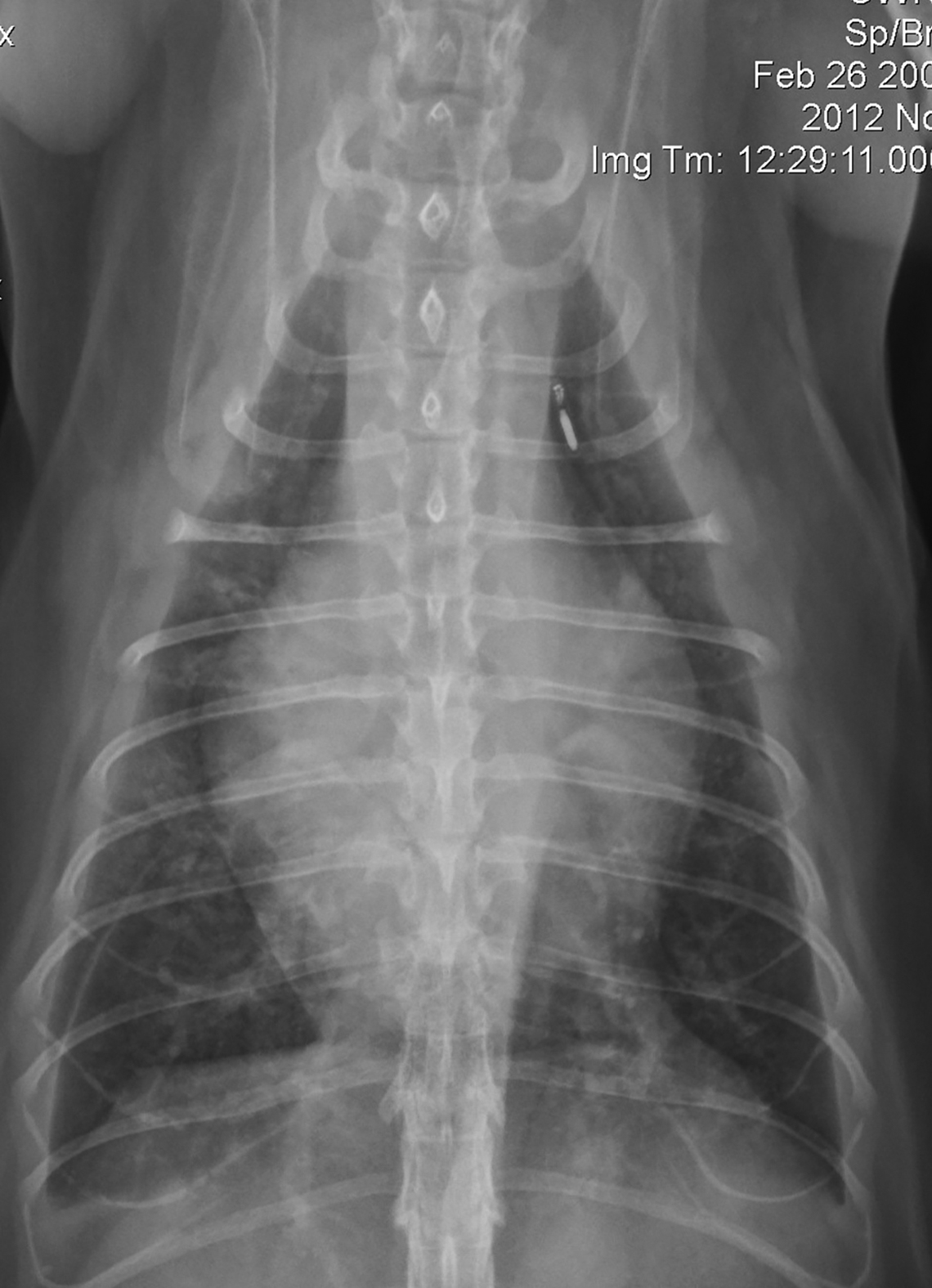
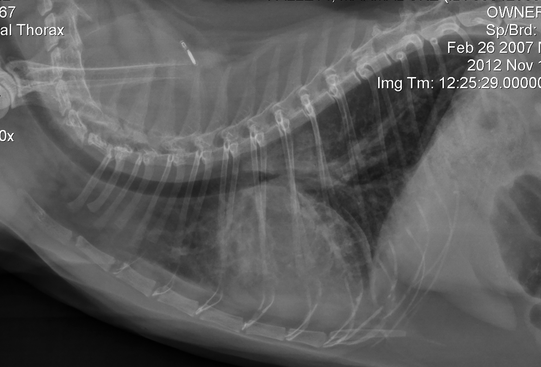
Radiographic interpretation The heart is obviously enlarged based on cardiac elongation and subjective analysis of shape heart on the DV projection. The VHS is still in the “gray zone”; i.e. suspicious for the cardiomegaly, but not definitive for CHF at this time. The left auricle is prominent on the DV and the left atrial outline, superimposed over the ventricles, is prominent (note splaying [*] of the main left and right bronchi; see the annotated DV image). There is some border effacement (obscuring) of the cardiac silhouette, especially ventrally on the lateral views compatible with pleural effusion. On the left lateral view the “down” caudal lung lobe is retracted slightly; compare this expansion to the right caudal lobe on the left lateral view (look below the “Feb” on the date label). On the DV projection, there are pleural fissure lines evident, especially on the right side (arrows on annotated image). Increased unstructured interstitial intrapulmonary densities are present and likely relate to pulmonary edema. These are patchy in distribution and might have been more widespread prior to diuretic therapy. Point-of-care ultrasound showed a small pleural effusion, pulmonary B-lines, and enlarged left atrium. Full echocardiography revealed end-stage (burned-out) hypertrophic cardiomyopathy (HCM).
Clinical interpretation/ additional case information This patient had a long-history of asymptomatic HCM (diagnosed years before) but was presented relatively acutely with clinical signs compatible with CHF. After initial hospital stabilization, Duke was treated with clopidogrel (anti-platelet therapy), and sent home with oral furosemide, pimobendan, enalapril. Spironolactone was eventually added to his chronic therapy. The smaller pills and cut pill fragments were combined in a #2 gel capsule to facilitate administration. Pimobendan is not labeled for cats, but is often used in feline CHF, especially in the absence of dynamic LV outflow tract obstruction. Thoracocentesis was not performed because the volume of effusion was relatively small. Duke was released to care of his owner but suffered recurrent bouts of CHF over the next 1-1/2 years requiring escalation of drug dosages.
Clinical History
Signalment: 5-year old MC Ragdoll cat
Clinical History Duke has a history of known heart disease and a systolic murmur and was presented for recent tachypnea and reduced appetite. The client indicated Duke’s resting respiratory rate increased from a consistent 28/minute to approximately 40/minute. On physical examination there was a faint systolic murmur, a prominent gallop sound, and premature beats detected. Duke was tachypneic and displayed increased respiratory effort. He was initially stabilized with butorphanol, oxygen, and 2-mg/kg of furosemide (IV) for a presumptive diagnosis of congestive heart failure. A point-of-care ultrasound exam was abnormal (details not shown). Radiographs were obtained a few hours after admission.