Era
Miniature Poodle
Select Radiograph(s)
Radiographic Report
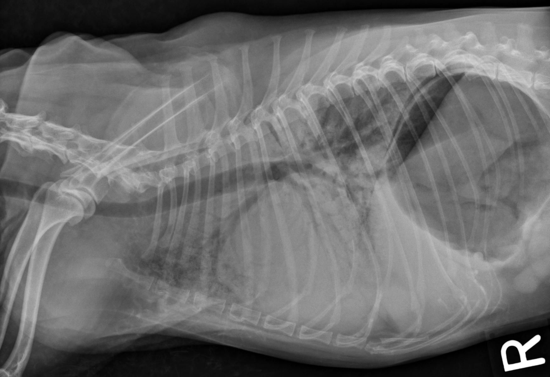
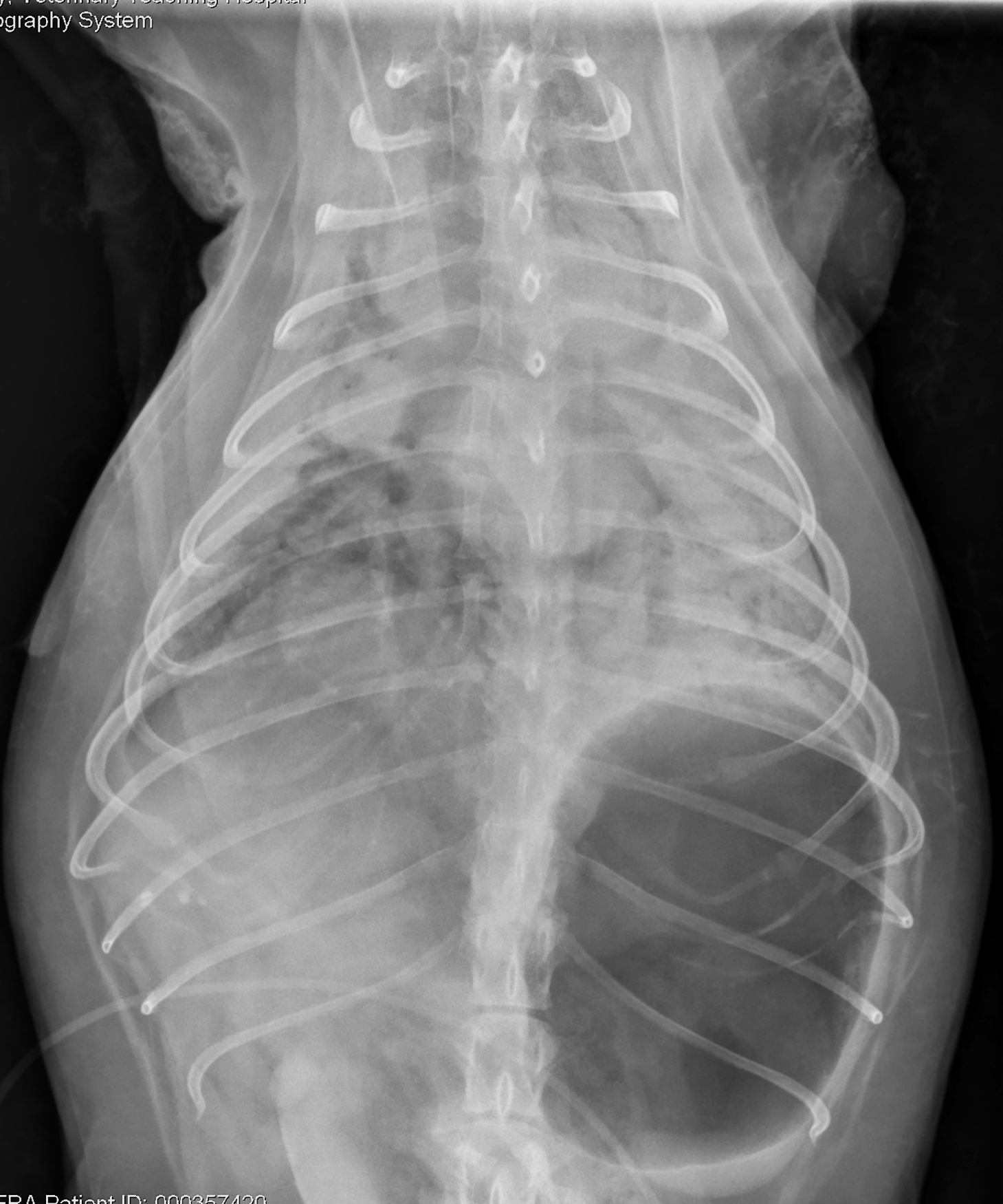
Radiographic interpretation: There is aerophagia due to respiratory distress. The heart cannot be seen clearly on the DV due to severe alveolar pulmonary infiltration. The lateral shows a cardiac silhouette that seems subjectively enlarged but with borders obscured (silhouetted) by the intrapulmonary infiltrates. The VHS and VLAS are at best approximations, as the cardiac borders cannot be clearly identified. Air bronchograms are evident on both views (also note the cranial lung lobes on the lateral view). These changes are typical of severe alveolar disease. The left atrium might be somewhat prominent on the DV (note bifurcation of mainstem bronchi) but the heart size could be normal for some breeds based on VHS. If the VLAS is accurate, the left atrium is enlarged. Vascular margins are not particularly clear.
Radiographic diagnosis: Radiographic findings are consistent with acute, severe left-sided congestive heart failure.
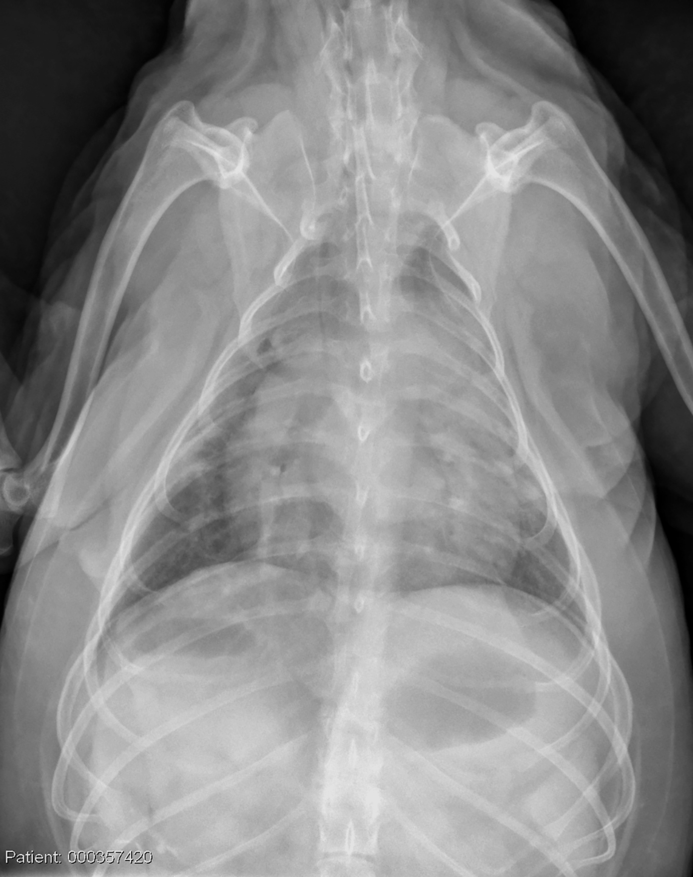
Clinical interpretation / additional case information: The clinical history and radiographs are compatible with fulminant left-sided CHF, but noncardiogenic reasons for edema or pulmonary hemorrhage cannot be ruled out. The history is typical of a ruptured chordae tendineae (flail leaflet), and this was evident on echocardiography. This dog was treated aggressively with IV furosemide (including a CRI), pimobendan, intravenous sodium nitroprusside, oxygen and sedation. Era responded amazingly well to treatment and a DV radiograph taken later that day showed significant improvement (single DV radiograph – 18h Post-treatment). The dog was released on oral medications. Dogs with flail leaflets often develop acute fulminant pulmonary edema; in these cases, cardiac size may be underwhelming but the edema can be explained by a large regurgitant volume entering a noncompliant left atrium. Reducing venous pressures, unloading the ventricles, and giving the heart time to remodel can allow some patients to be stabilized and released.
Clinical History
Signalment: 10 year old FS miniature poodle
Clinical History: Era was presented emergently for acute onset of respiratory distress. She was normal in the morning and was in respiratory distress when the owner returned from work. The patient had been confined all day and there was no evidence of trauma. She had a prior history of a cardiac murmur without overt signs other than occasional coughing. Era was presented to the emergency department with respiratory distress, effort, and a breathing rate of approximately 100 breaths/minute. Her pulse rate was 90 and regular. She expectorated blood-tinged foam. The heart could not be heard; diffuse crackles and wheezes were evident. After sedation and an injection of furosemide, DV and right lateral radiographs were obtained.