Flower
Mix Breed
Select Radiograph(s)
Radiographic Report
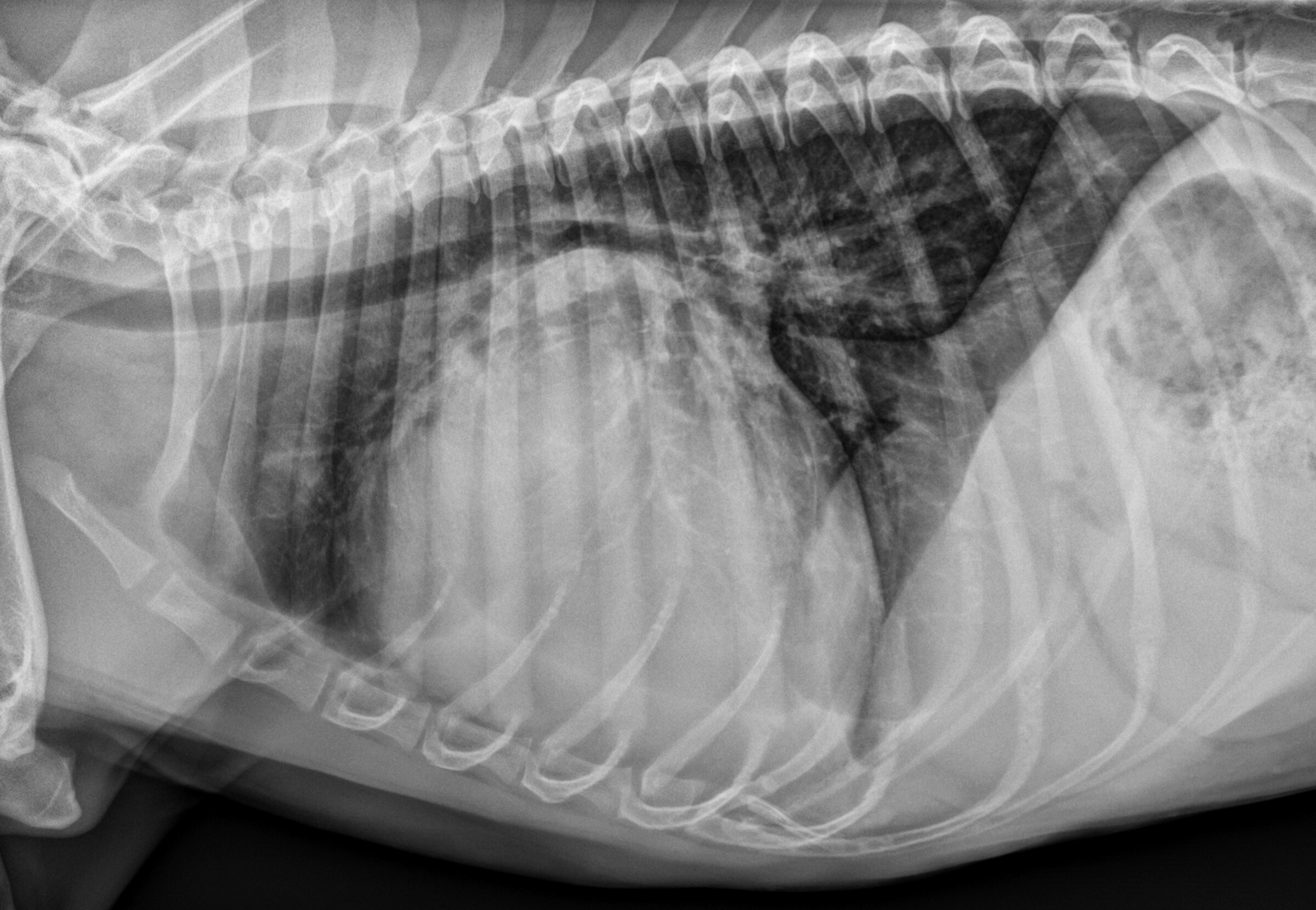
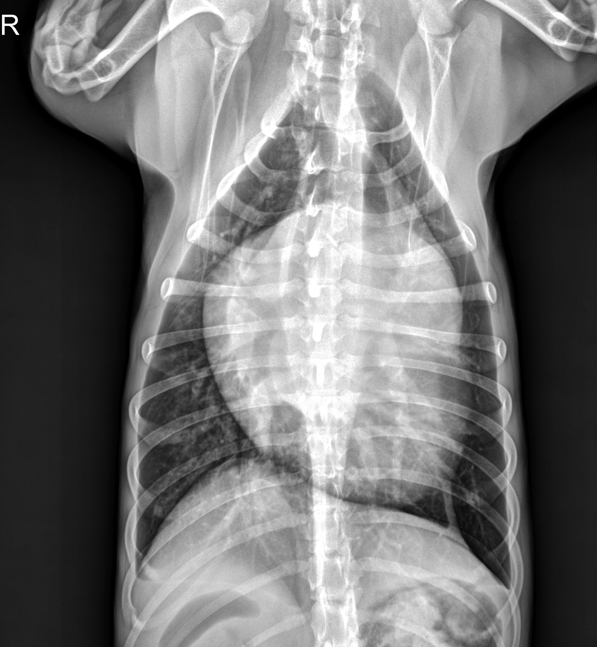
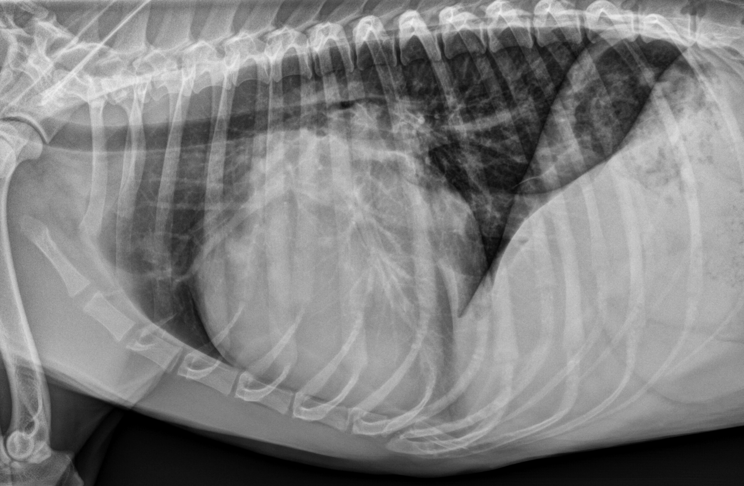
Radiographic interpretation: The heart is generally enlarged with dorsal deviation of the trachea. There is moderate left atrial enlargement with splaying of the principal bronchi and double soft tissue opacity sign on the VD image. There is a cardiac bulge at the 2-3 o’clock position on the VD image, consistent with enlargement of the left auricular appendage. There is increased sternal contact on the lateral images, suggesting enlargement of the right ventricle. A cardiac bulge is seen at the 9-11 o’clock position on the VD image, consistent with right atrial enlargement. There is a mild interstitial pattern in the parenchyma on the left lateral image. There is a moderate bronchial pattern in the caudal lung lobes. The cranial pulmonary vein on the left lateral image is enlarged compared to the corresponding artery. The mediastinum and diaphragm are normal.
Radiographic diagnosis: The enlarged cardiac silhouette supports a cardiogenic cause for the respiratory distress with congenital heart disease or dilated cardiomyopathy being most likely. The mild interstitial pattern and diffuse bronchial pattern suggest mild pulmonary edema. These findings together with dilation of the pulmonary veins supports a diagnosis of left-sided congestive heart failure (CHF).
Clinical interpretation/additional case information: On Sophie’s echocardiogram, severe four-chamber dilation was apparent with very reduced systolic function (fractional shortening of 11%). The client reported that the dog had been eating a boutique, grain-free diet for most of the dog’s life. Nutritional cardiomyopathy was considered possible and taurine levels were submitted. Given the low blood pressure and concern for CHF, she was hospitalized in any oxygen cage with 40% oxygen and received IV furosemide as well as oral pimobendan and taurine. The next morning her respiratory rate had declined to 30 breaths per minute and her heart rate was now 130bpm. She was discharged receiveing furosemide, pimobendan, and taurine with recommendations to change to a commercial non-grain free diet.
Clinical History
Signalment: 3 year old FS mixed breed dog
Clinical history: The patient was presented to an urgent care for respiratory distress and inappetence; she has also developed a progressive cough over the past month. Over the prior 2 days she was uncomfortable with a noticeable abdominal component to her breathing, especially while laying down. She is also exhibiting signs of exercise intolerance and is unwilling to walk far on her daily walks. On physical examination, she was tachycardic (200 bpm) and had femoral pulse deficits. A grade II/VI left apical systolic heart murmur was appreciated. Her respiratory rate was 70 breaths per minute with increased effort but no appreciable crackles. Systolic blood pressure performed on the left rear limb was 85 mmHg.