Flutter
Domestic Shorthair
Select Radiograph(s)
Radiographic Report
Radiographic interpretation:
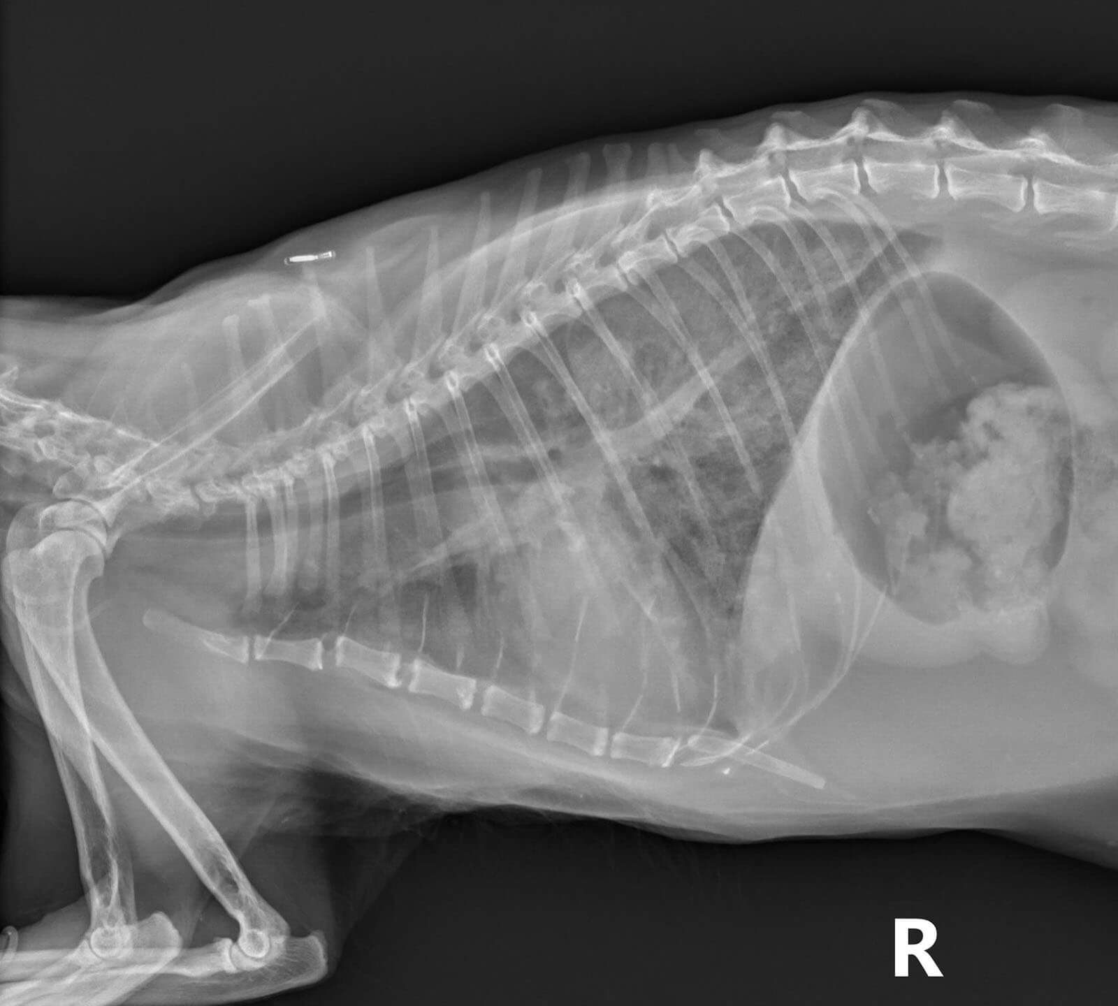
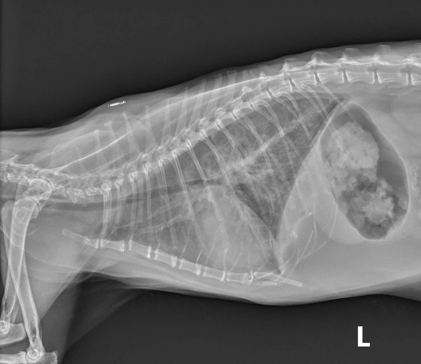
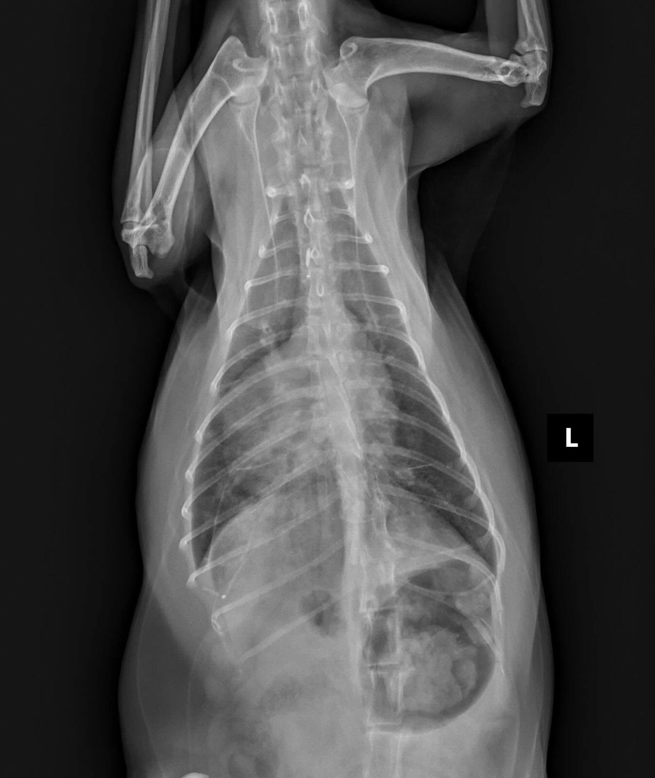
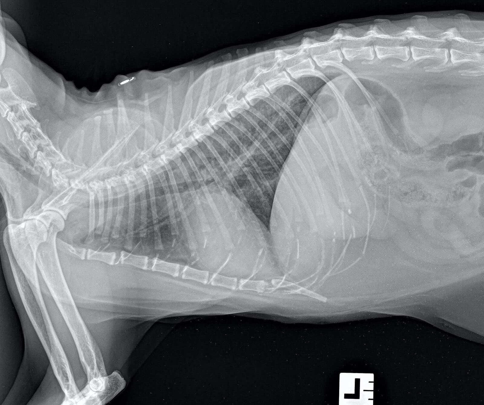
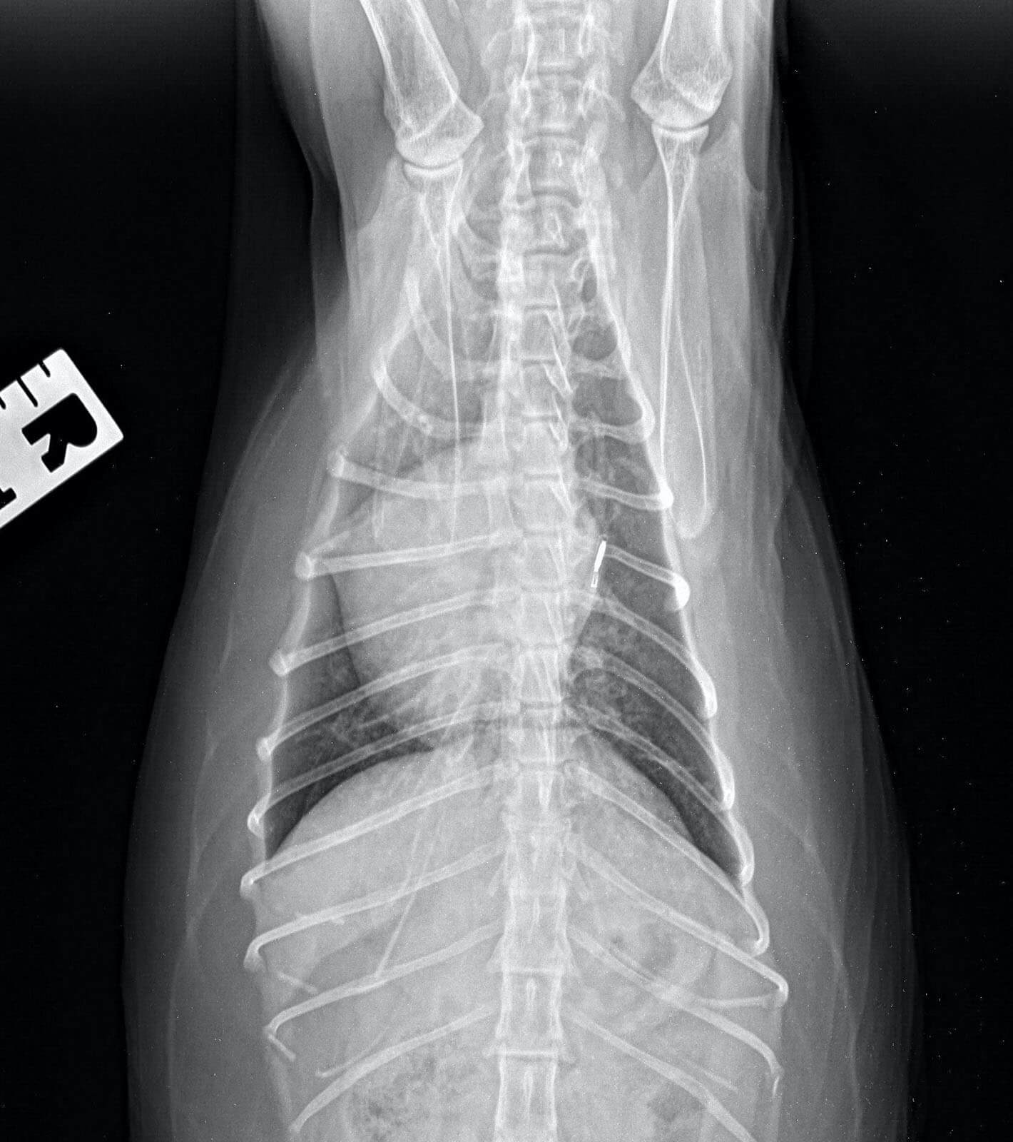
Visit 1: Moderate to severe cardiomegaly with possible left atrial dilation noted on the VD view, but the patient alignment is not optimal. There is severe pulmonary venous distention and the margins of the pulmonary veins and arteries, both caudal and cranial to the heart, are obscured. There is a caudal dorsal interstitial pattern with some pleural effusion suspected. Both of these are more severe in the right caudal lung and thorax. Echocardiographic results confirm eccentric hypertrophy of all four chambers.
Radiographic diagnosis: Cardiomegaly and findings consistent with congestive heart failure.
Clinical interpretation / additional case information: Radiographic, echocardiographic and laboratory results are consistent with CHF secondary to thyrooxicosis. Furosemide and enalapril were administered and the patient was switched to oral methimazole.
Radiographic interpretation:
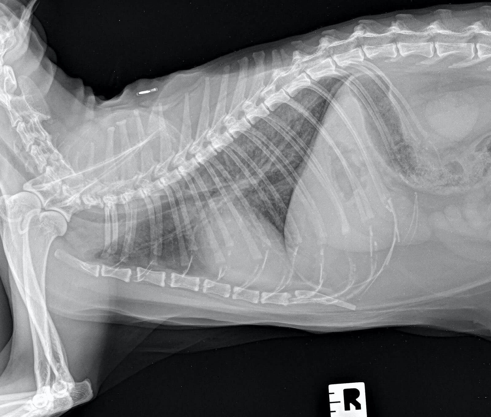
Visit 2, 7 days post treatment: Significant reduction in heart size (VHS=8.8) and improvement in the interstitial pattern, pulmonary venous distention and vascular detail. There is still a bronchial pattern remaining in the caudal lung lobes.
Clinical History
Signalment: 14yr old MC Domestic Shorthair
Clinical History: Patient was previously diagnosed with hyperthyroidism and is being treated with methimazole administered via transdermal gel once daily. The recent total T4 was 6.2mg/dl. The patient was found under the bed today, tachypneic with increased respiratory effort. The family rushed him to the veterinary emergency hospital. Upon presentation, a gallop heart sound was detected and bilateral pulmonary crackles were noted. 2 sets of radiographs are presented.
