Maggie
Shih Tzu
Select Radiograph(s)

Radiographic Report
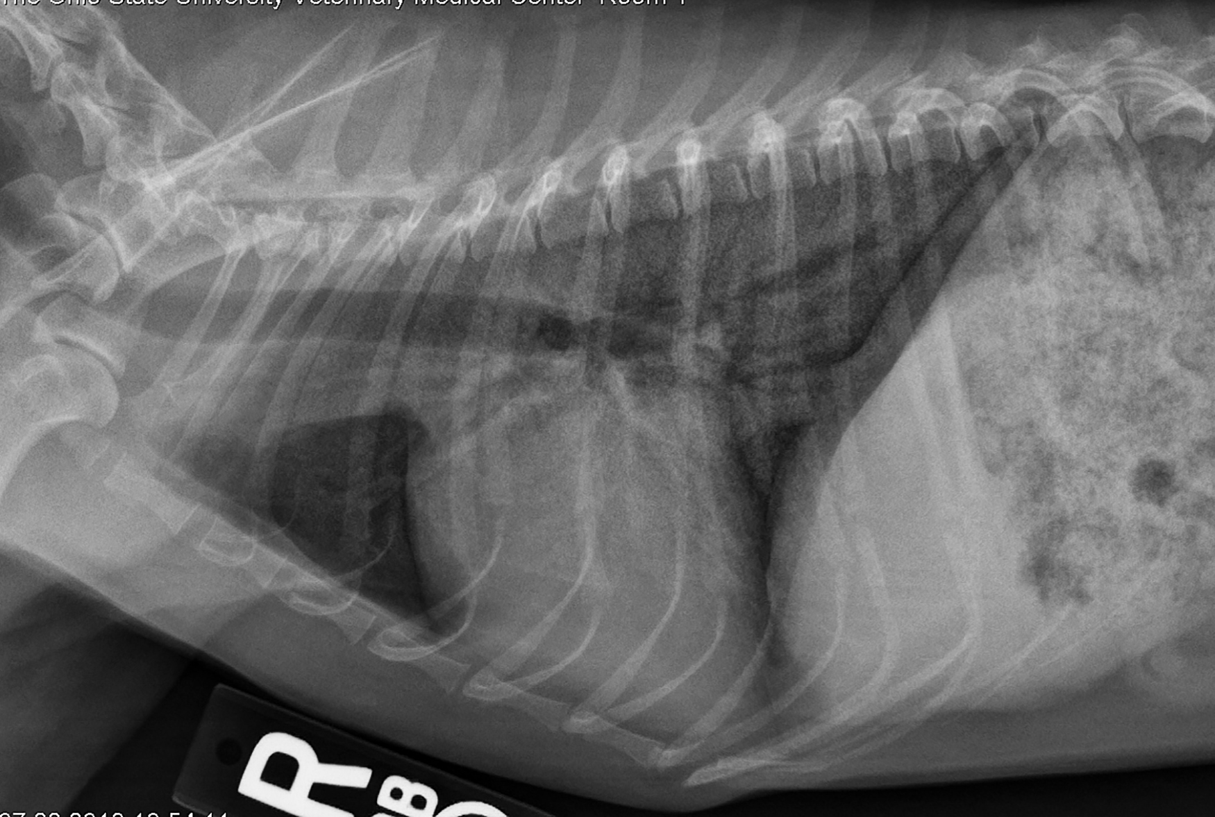
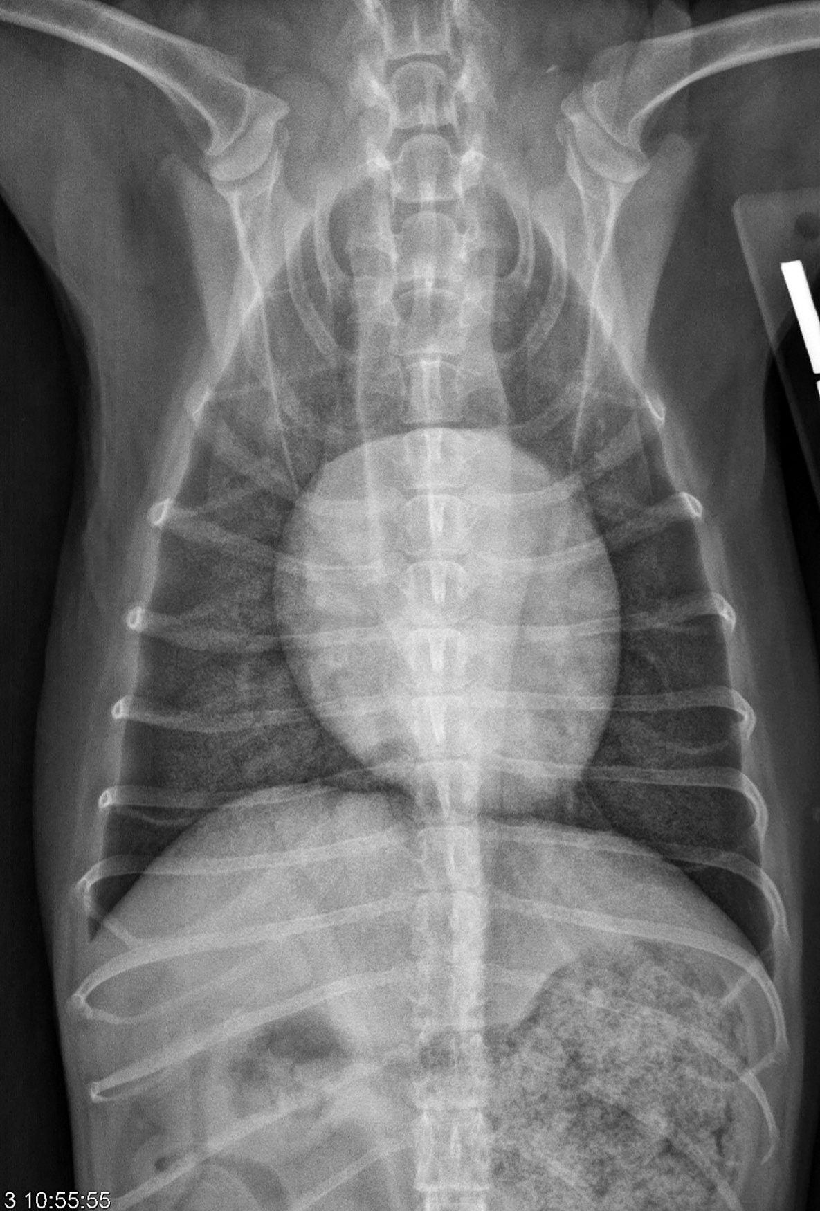
Radiographic interpretation: The heart is not significantly enlarged based on VHS, LAS or subjective evaluation. The left lateral (as often is the case) shows mild apex elevation, a slightly more prominent left atrium, and slightly larger cranial pulmonary vein (compared to artery); these are not evident on the right lateral. The major abnormality is a diffuse increase in unstructured interstitial lung density involving all lung lobes. The lungs exhibited an almost granular appearance, typical of calcification. Airways are visible in distal lung fields suggesting alveolar involvement or pulmonary densities sufficient to outline the airways. Whether this is true “alveolar” fluid infiltration, or simply outlining of larger airways by a dense lung parenchyma is uncertain, but the latter is most likely.
Clinical interpretation/ additional case information: Based on the history, examination findings, and an echocardiogram that showed only mild myxomatous mitral valve disease and regurgitation, a diagnosis of probable pulmonary fibrosis with mineralization was made. This is almost certainly iatrogenic, related to the 9 years of continual prednisone administration and similar to spontaneous cases of severe, untreated Cushing’s-like disease in dogs. Although cardiogenic and noncardiogenic pulmonary edema were considered, the dog was not in respiratory distress and the cardiac and vascular findings did not support that diagnosis. Cardiac medications were not prescribed; the changes in the lung were considered irreversible, and the owners were advised to lower body weight (to limit thoracic restriction) and control exercise for their dog. No treatments have been shown to be effective for pulmonary fibrosis and because echocardiography did not indicate severe pulmonary hypertension, sildenafil was not prescribed. The patient was returned to the care of the referring DVM for management of iatrogenic hyperadrenocorticism and serial radiographs were recommended to follow the course of mitral valve disease.
Clinical History
Signalment: 10-year old FS Shih Tzu
Clinical History: Maggie was referred for chronic and progressive rapid breathing (tachypnea) as well as exercise intolerance. She has a long history of “liver disease” that was diagnosed by another veterinarian. This disorder had been treated with prednisone for an elevated serum ALT concentration for nearly nine years (!). Their current family veterinarian had detected a grade 2-3/6 systolic murmur loudest over the left apex and bilateral lung crackles. Hyperpigmentation of the skin and symmetric hair thinning were also noted dorsally. Serum biochemical tests showed a mild increase in ALT and AST and severe elevation in serum alkaline phosphatase. Current medications included heartworm prevention and prednisone, now given every other day and at decreasing dosages under the direction of their current (referring) veterinarian. On referral examination our findings were as reported by the referring veterinarian. An echocardiogram and thoracic radiographs were obtained.