Matchu
DSH
Radiographic Report
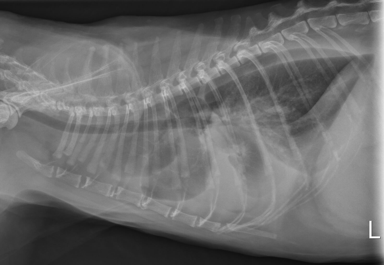
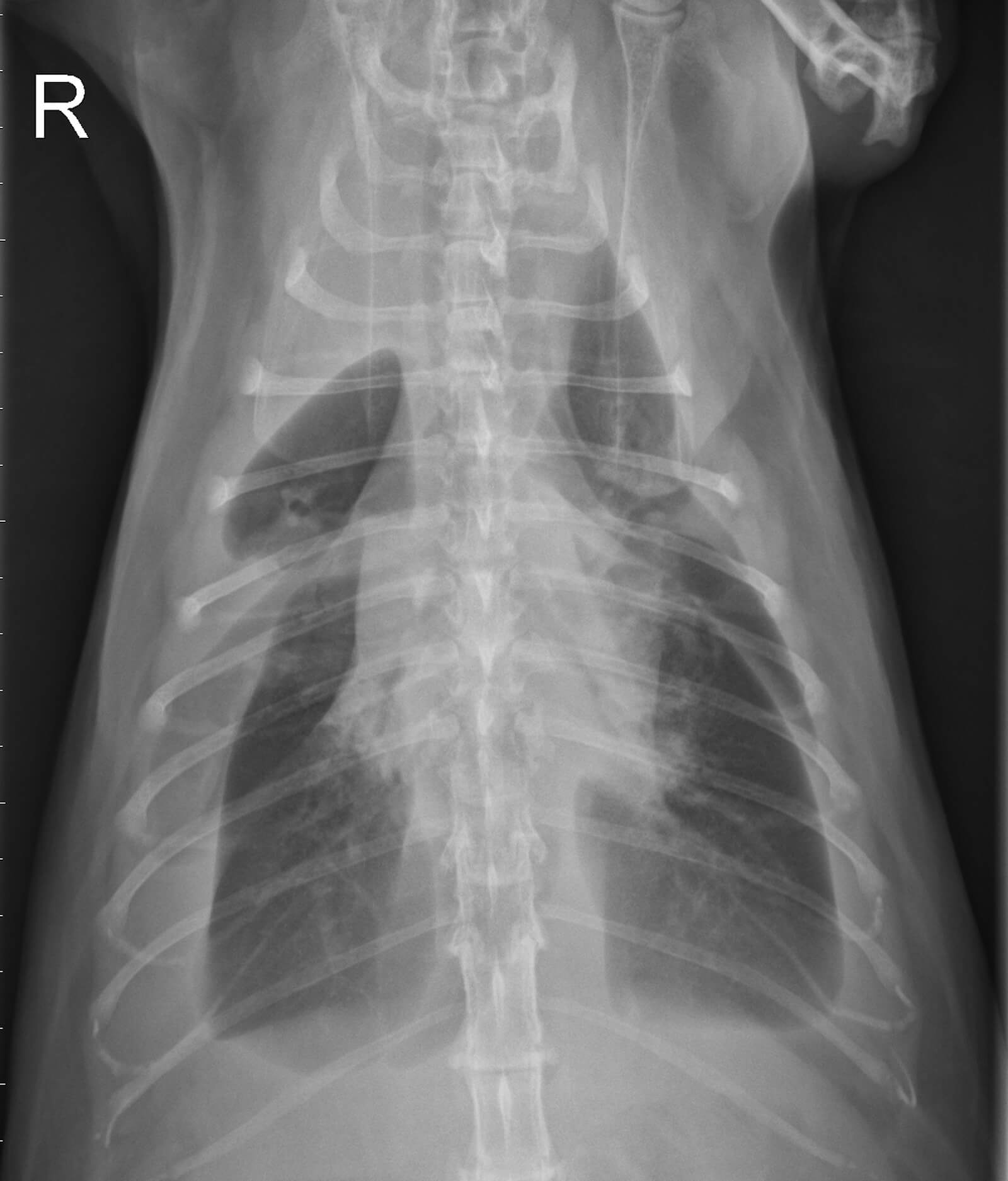
Radiographic interpretation The cardiac silhouette is obscured by moderate to severe pleural effusion. Vertebral heart scale cannot be calculated, but the heart appears to be enlarged, especially on the lateral view. The lung lobes are partially retracted and appear to be rounded, which may be an indication of pleural fibrosis.
Clinical interpretation/additional information: The differential diagnosis list for feline pleural effusion includes congestive heart failure, chylothorax, pyothorax or neoplastic effusions. In this cat, the presence of distended jugular veins on physical examination supports a cardiac origin for the effusion (i.e. congestive heart failure). After thoracocentesis and stabilization, echocardiographic examination revealed restrictive cardiomyopathy. The rounded appearance of the lung lobes may reflect pleural fibrosis, and in this cat, likely indicates that the onset of effusion was more chronic than it appeared based on clinical signs.
Clinical History
Signalment: 11 year old MC DSH
Clinical history: Matchu was presented for acute onset of difficulty breathing. On physical examination, the patient was tachypneic, dyspneic and had a heart rate of 220 bpm with a regular rhythm and poor femoral pulses. Jugular distension was noted.

