Max
Boxer
Select Radiograph(s)
Radiographic Report
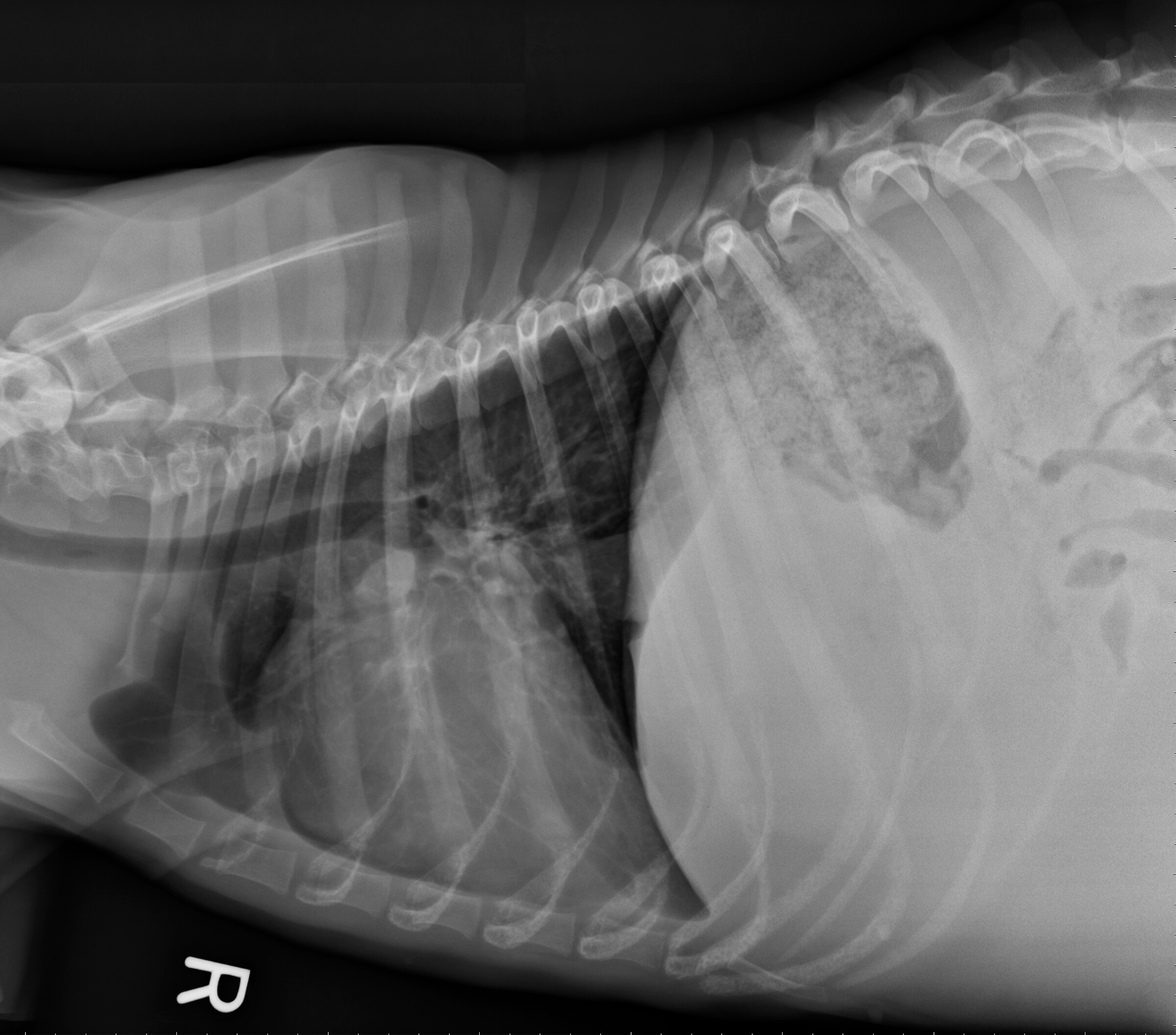
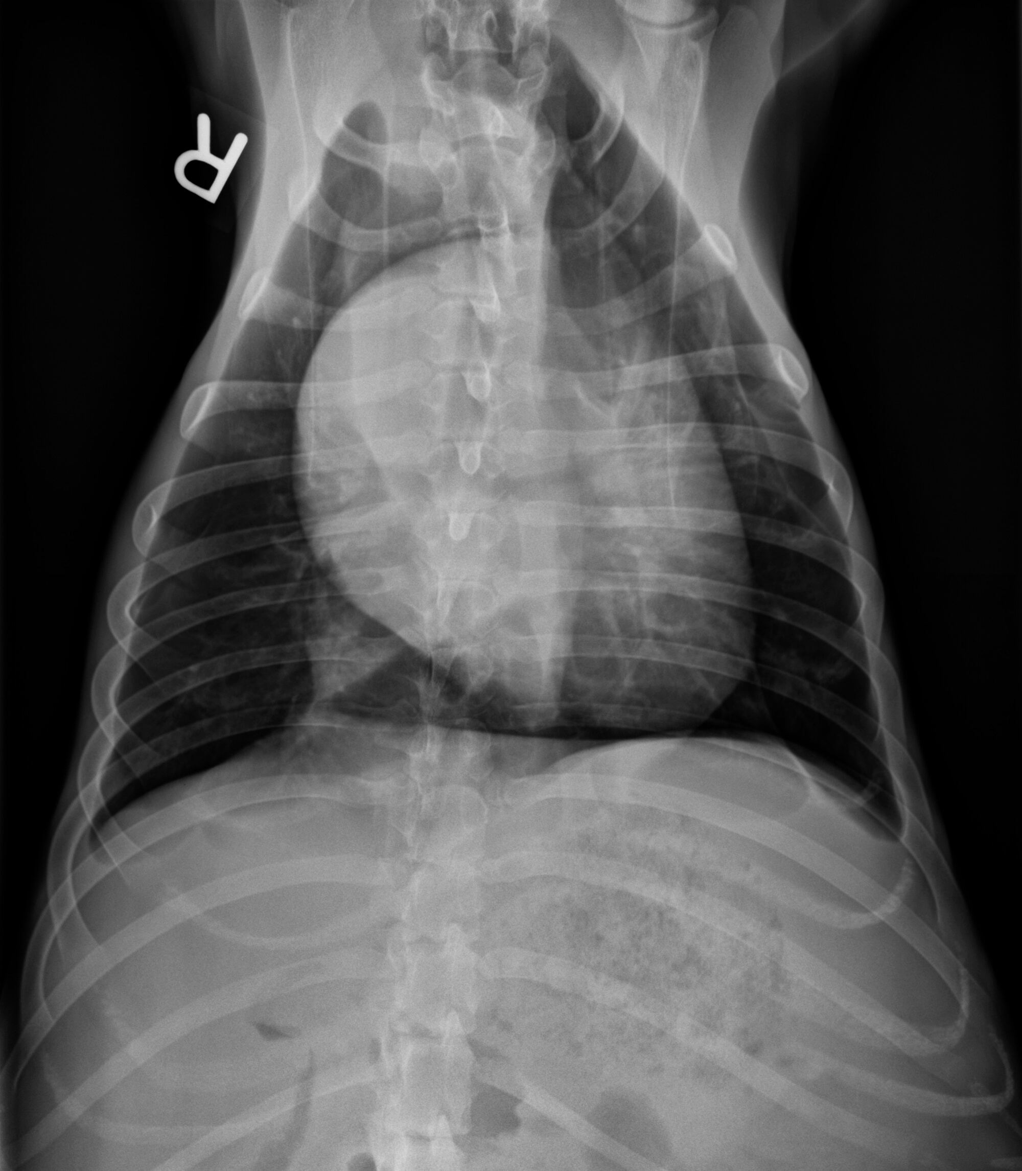
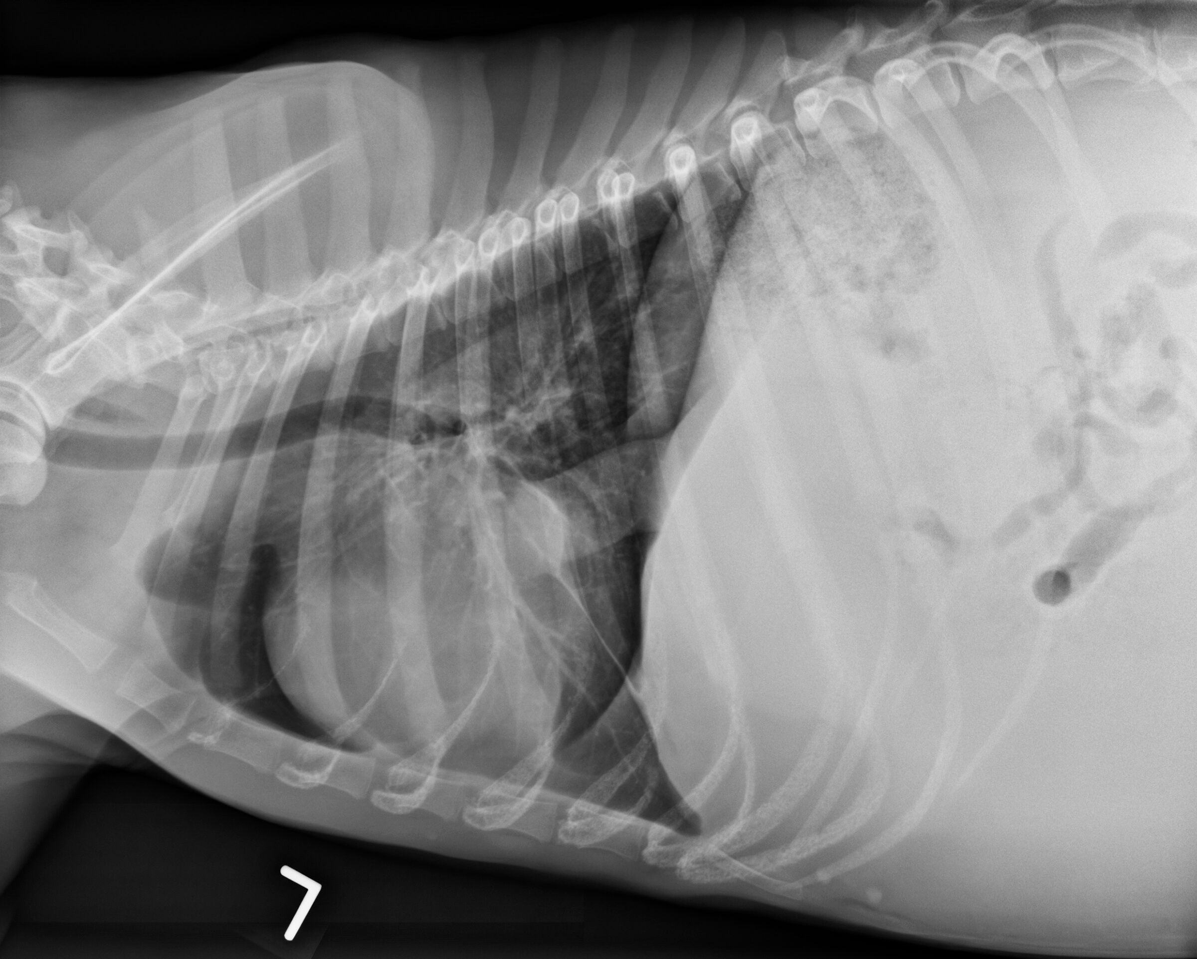
Radiographic interpretation: There is severe right atrial enlargement. Slight pleural fluid effusion is apparent around the right caudal, accessory, and left lung lobes. The caudal vena cava is markedly distended. There is loss of abdominal detail consistent with severe ascites.
Radiographic diagnosis: The radiographic findings are consistent with right-sided heart disease and right-sided congestive heart failure (CHF).
Clinical interpretation/additional case information: Echocardiographic examination showed severe pulmonary valve stenosis as well as tricuspid valve dysplasia. These two congenital defects in combination can lead to progressive right heart enlargement and the eventual development of right-sided congestive heart failure. Treatment included therapy for CHF – furosemide, enalapril, pimobendan, spironolactone – as well as diltiazem therapy for the atrial flutter.
Clinical History
Signalment: 2 year old MC Boxer dog
Clinical history: Max has a history of a heart murmur identified at 8 weeks of age. He began having syncopal events at 6 months of age, occurring during periods of excitement. The owners have recently noticed that Max has abdominal distension, which has become marked over the past 4 weeks. On examination, there is a grade III/VI left basilar systolic heart murmur with a rapid and irregular heart rhythm (diagnosed by ECG as atrial flutter with variable conduction); pulse quality is irregular with deficits. The jugular veins are distended. The abdomen is distended with a palpable fluid wave.