Minnie
Cavalier King Charles Spaniel
Radiographic Report
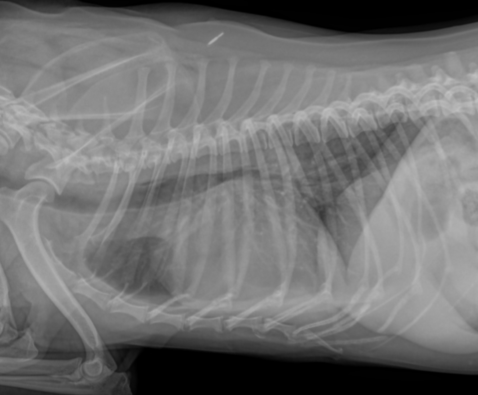
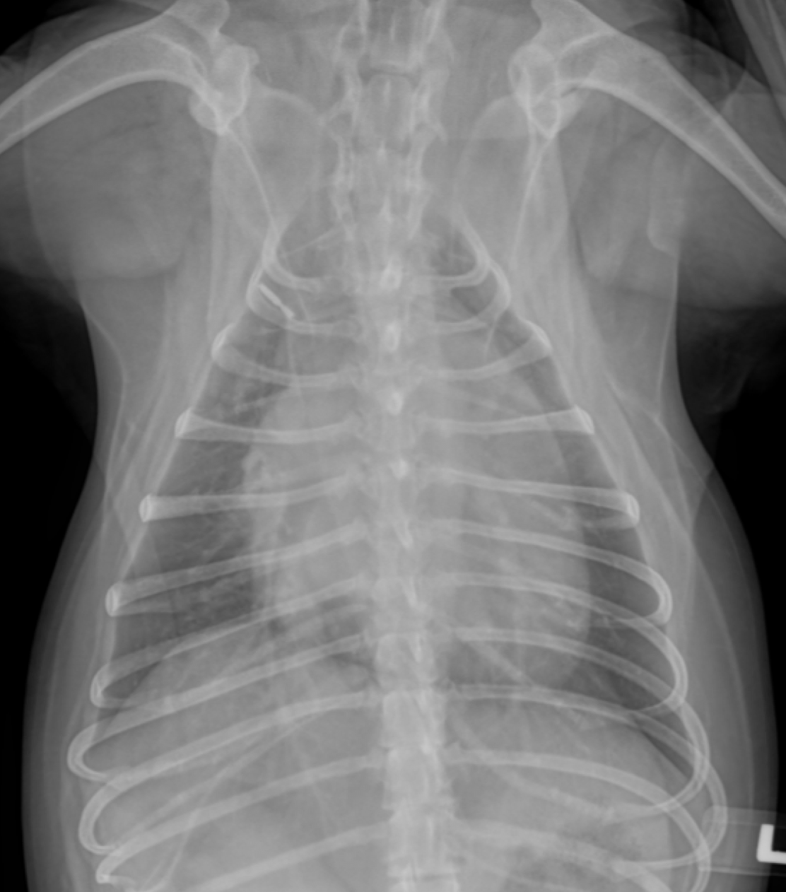
Radiographic interpretation: The cardiac silhouette is enlarged with a VHS of 12.3 and a VLAS of 2.9. Obvious left atrium and left ventricle enlargement is present. There is a mild increase in nonspecific interstitial opacities, but no overt evidence of pulmonary edema is appreciated. Pulmonary vessels are difficult to assess; the mainstem bronchi appear to be compressed as a consequence of the enlarged heart.
Radiographic diagnosis: Enlarged cardiac silhouette with no evidence of active CHF; airway compression is evident.
Clinical interpretation/ additional case information: An echocardiogram is recommended to better define the nature and extent of the heart disease, but given the degree of changes on the thoracic radiographs coupled with a typical breed and murmur for MMVD, this patient is suspected to have Stage B2 MMVD and treatment with pimobendan (+/- ACEi) is reasonable based on the films alone. In the absence of CHF, the addition of a cough suppressant is also reasonable if the cough is deemed clinically significant.
Clinical History
Signalment: 10 year old FS Cavalier King Charles Spaniel
Clinical History: Presented for cough with one documented syncopal episode following a coughing spell. A loud (4/6) left apical systolic murmur was present.