Notre Dame
Radiographic Report
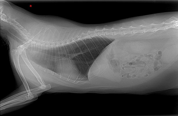
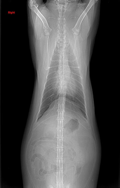
The films are of good technical quality, but could optimally be collimated to exclude the abdomen. The VHS measures 8.0 and the cardiac silhouette appears to be somewhat globoid and enlarged, but no specific chamber enlargement is identified. There is, however, a ring of a slightly lighter radiographic density around the silhouette, most pronounced over the left ventricle and left atrial appendage, but also present on the right side, a density consistent with adipose tissue overlying a soft tissue density (cardiac silhouette). There is no pulmonary venous congestion nor pulmonary arterial distension. A mild pulmonary infiltrate is diffusely distributed with a very mild diffuse bronchial pattern.
Further Testing: An echocardiogram was performed and revealed normal cardiac size with pericardial, epicardial, and sternal fat. The interpretation was normal cardiac structures with excessive epicardial and pericardial fat.
Treatment/Management: Reduce caloric intake.
Clinical History
Notre Dame is a 9-year-old male neutered domestic shorthair. The client describes mild exercise intolerance and he has gained 3 lbs over the past 12 months. The patient has no previous history of cardiopulmonary disease.