Peanuts
DSH
Radiographic Report
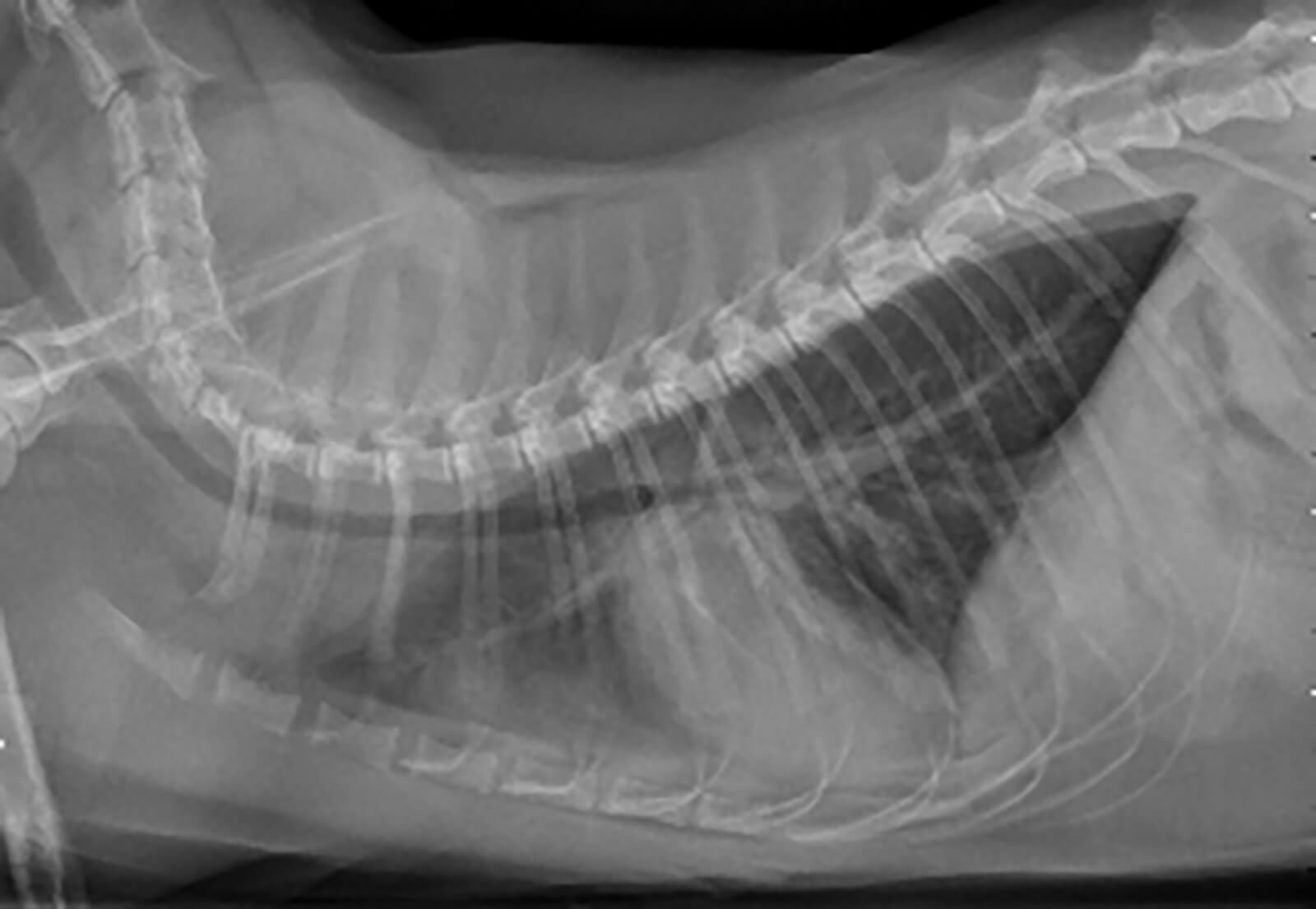
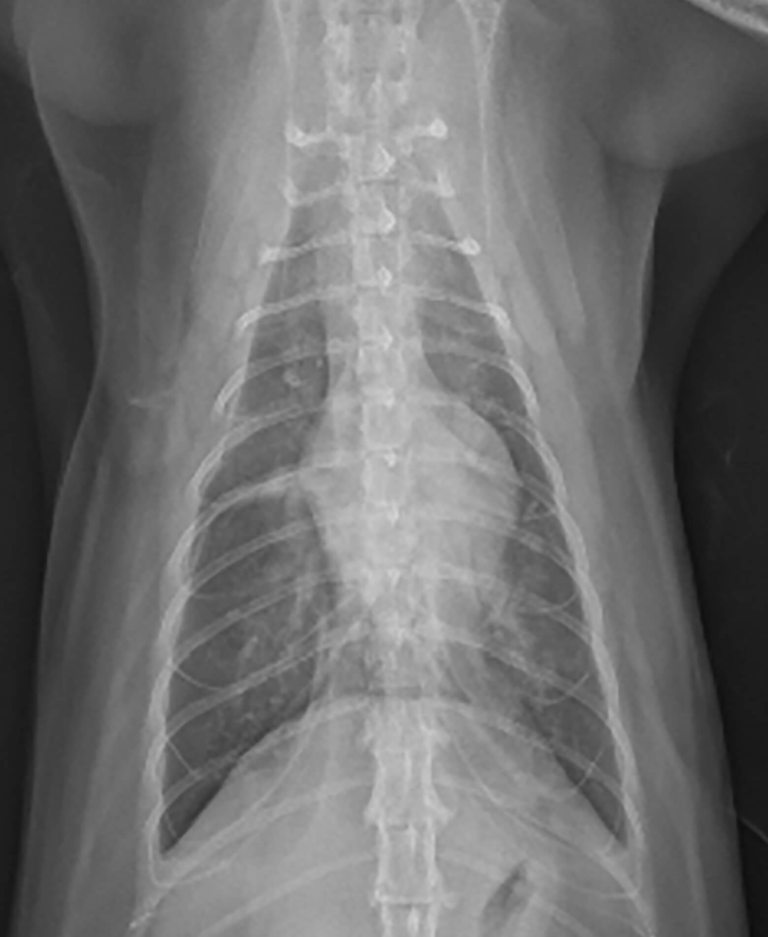
Radiographic interpretation: VHS 8.6 (moderate cardiomegaly), elongated cardiac silhouette on lateral view indicative of left ventricular enlargement. On VD view, the left atrium appears to be enlarged (open arrow on annotated VD image). Alveolar infiltrates are present in the cranial ventral and caudal ventral pulmonary fields on the lateral view, and can be seen in the caudal fields bilaterally (small arrows on annotated VD view). The pulmonary arteries and veins are enlarged, and a small amount of pleural effusion can be seen “outlining” the caudal dorsal lung lobes.
Radiographic diagnosis: The combination of cardiomegaly, alveolar infiltrates and enlargement of pulmonary arteries and veins in a dyspneic feline patient is suggestive of congestive heart failure. In this patient, only a small amount of pleural effusion is noted, but pleural effusion is a common finding in cats with congestive heart failure. Echocardiographic examination in this patient revealed the presence of hypertrophic cardiomyopathy.
Clinical History
Signalment: 4 year old MC DSH
Clinical history: 2 day history of not eating and drinking, and hiding under the bed. The owners noticed that he has started to breathe more heavily in the past 24 hours.