Penny
Domestic Shorthair
Radiographic Report
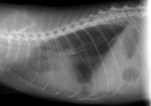
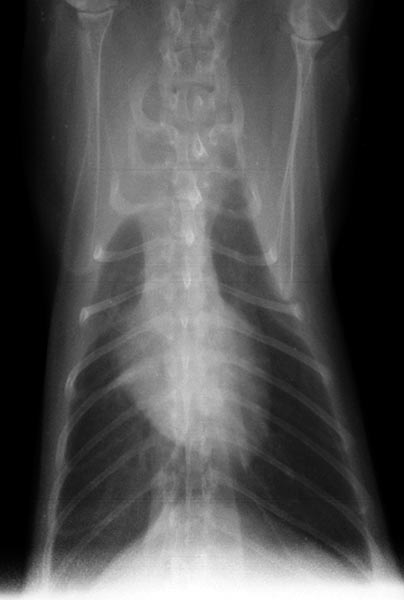
The heart is subjectively enlarged, appearing tall on the lateral view, but is obscured in part by fluid density and cannot be completely assessed. Careful inspection of the caudal lung fields on the VD shows a retraction of the borders, indicative of pleural effusion bilaterally. Pleural effusion is also evident on the lateral view. There is border effacement (silhouetting) of the cardiac and diaphragmatic shadows with a widened cranial mediastinum. The cranial lobes are displaced caudally (see the lateral view), especially the right cranial lobe (see the VD view). Additionally, on the lateral view, there is the typical scalloped appearance of a fluid/air interface in the ventral 1/3 of the thorax. The caudal vena cava appears very dilated, though somewhat obscured by pleural or mediastinal fluid. Pulmonary vessels appear normal in size. The diaphragm appears intact. On the lateral view the caudal blood vessel margins are clear, suggesting there is no significant intra-pulmonary density. Radiographic interpretation: Pleural effusion in cats is a common problem and can be caused by congestive heart failure, neoplasia (lymphoma, metastatic disease), chylothorax, FIP, pyothorax, and hemithorax among other causes.
With pleural effusion, the borders of the heart cannot be accurately identified on the right lateral view. Consequently VHS cannot be calculated.
Clinical History
Penny is a 7 year old FS DLH that presented for evaluation of tachypnea noted by the referring veterinarian. The cat has a poor appetite and is less active. Physical examination also indicated prominent jugular veins and a possible gallop sound.