Queeny

Radiographic Report
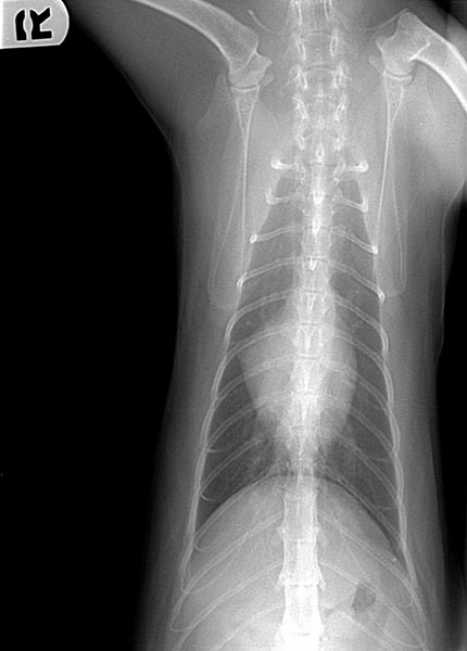
Technical Details Location of images: Thoracic radiographs obtained. Views of images: Left lateral and ventrodorsal throracic radiographs.Radiographic Findings Technical issues: None. Cardiac size including VHS: VHS 7.0. Cardiac silhouette appear mildly elongated, but this is suspected to be fat in the pericardiacophrenic ligament; the VHS is measured so that is not included. There is also an opacity at the cranial base of the heart that is most commonly the ascending aorta/aortic arch, but can be the right auricle or pulmonary artery. Cardiac silhouette appears to have a normal size, except for a slight opacified bulge near the right atrium. This is generally a difficult area to interpret, as this could be adipose tissue, but given the mildly dilated pulmonary arteries, right atrial dilation must be a consideration. No pulmonary venous congestion, but the pulmonary artery to the cranial lung field (lateral) and to the right caudal lung field (VD) appear mildly distended.Pulmonary infiltrate distribute predominately perihilar and caudodorsal. Pulmonary infiltrate pattern, bronchial. No pleural effusion. Other Findings: None. Treatment/Management: Patient was placed on a 3 week tapering dose of prednisolone 2.5mg every 12 hours. An albuterol metered dose inhaler was prescribed with an Aerokat(r) feline aerosol chamber if needed for asthmatic attacks leading to severe coughing or wheezing. Inasmuch as airway inflammation underlies asthma, the need for long-term anti-inflammatory therapy was discussed including using inhaled fluticasone if there is a good response to oral prednisolone. Clients were advised to view Youtube(r) videos regarding use of an inhaler. The medication costs associated with inhaled fluticasone were also mentioned, as this can be a practical factor affecting the acceptance of that treatment.
Clinical History
This feline Patient presented for frequent coughing and wheezing. Patient has a history of chronic cough as well as severe, recurrent skin and ear infections suspected to be secondary to allergies. The cat has slightly prolonged respiratory phase but no distress. No murmurs or gallops were detected.