Roo
Rottweiler
Select Radiograph(s)
Radiographic Report
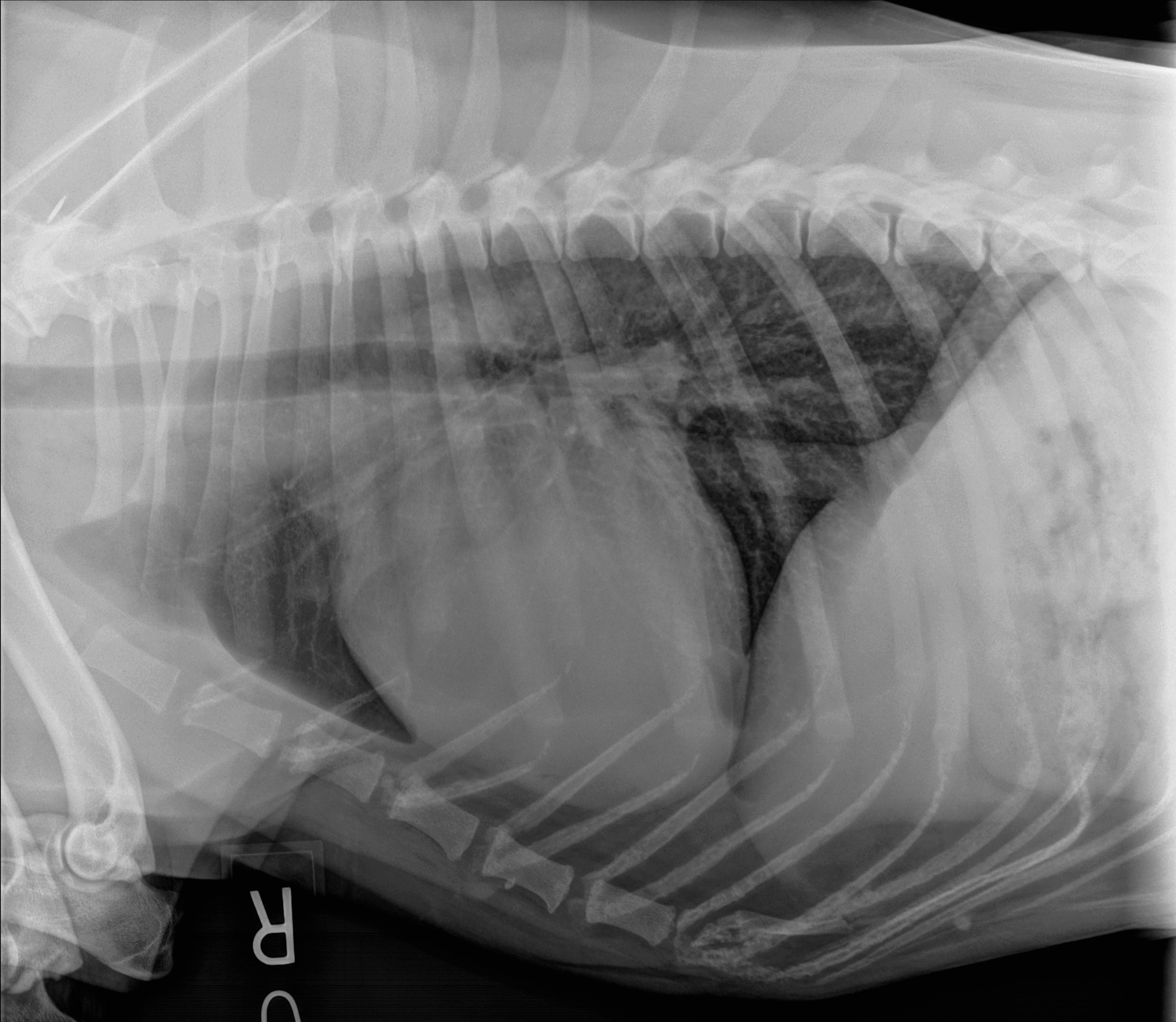
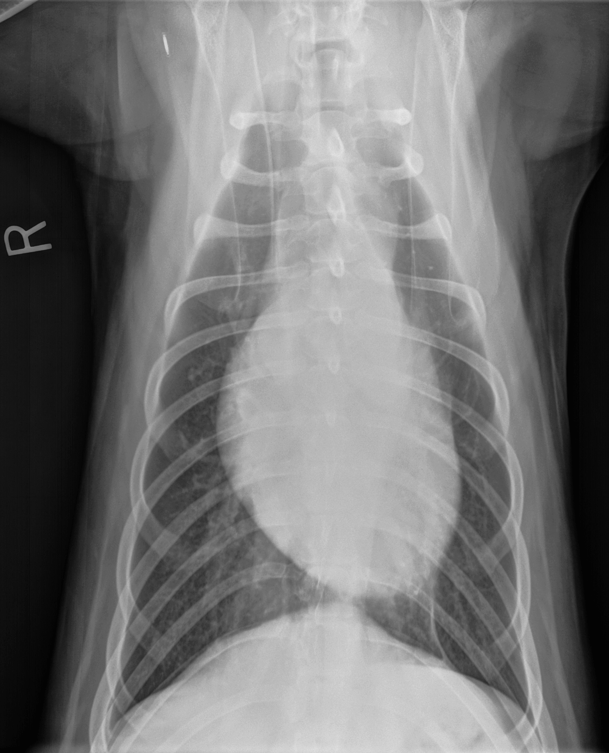
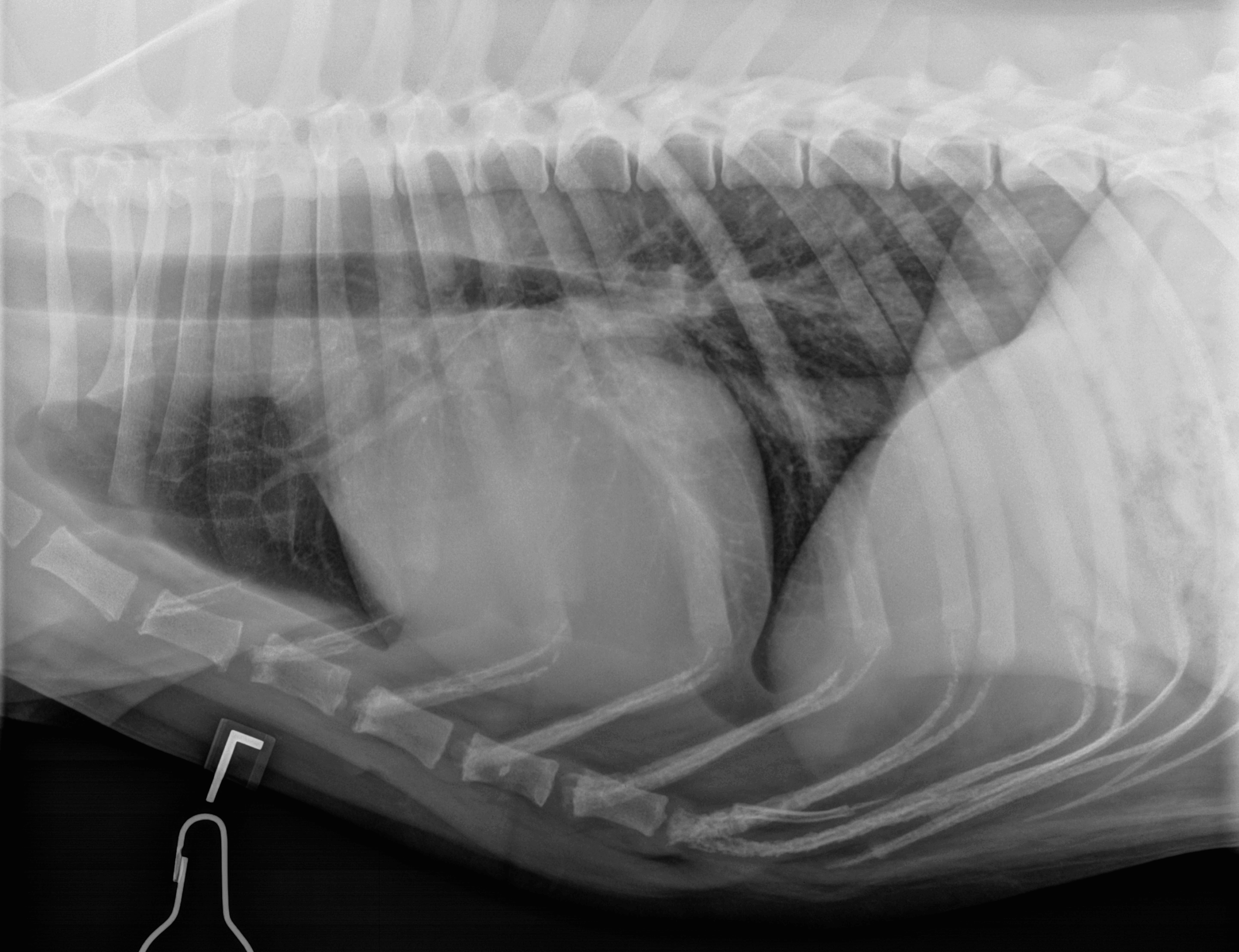
CEG Interpretation: Three views of the thorax were reviewed, comprising a complete thoracic study. The films demonstrate appropriate positioning and exposure. The cardiac silhouette is mildly enlarged on the right lateral view (VHS 10.8) with no evidence of left atrial enlargement (VLAS 2.0). The pulmonary parenchyma is clear, and pulmonary vascular markings are considered normal in size. There is no evidence of pleural effusion. No significant skeletal abnormalities are noted. The reported normal VHS range for the Rottweiler breed is 9.6-10.1
Clinical History
Signalment: 1-year-old, F, Rottweiler (36.0 kg)
The dog was presented for annual health evaluation. According to the owner the dog is apparently healthy . Auscultation revealed a moderately loud left apical systolic heart murmur (3 to 4 /6). Thoracic radiographs were performed.