Rufuss
DSH
Radiographic Report
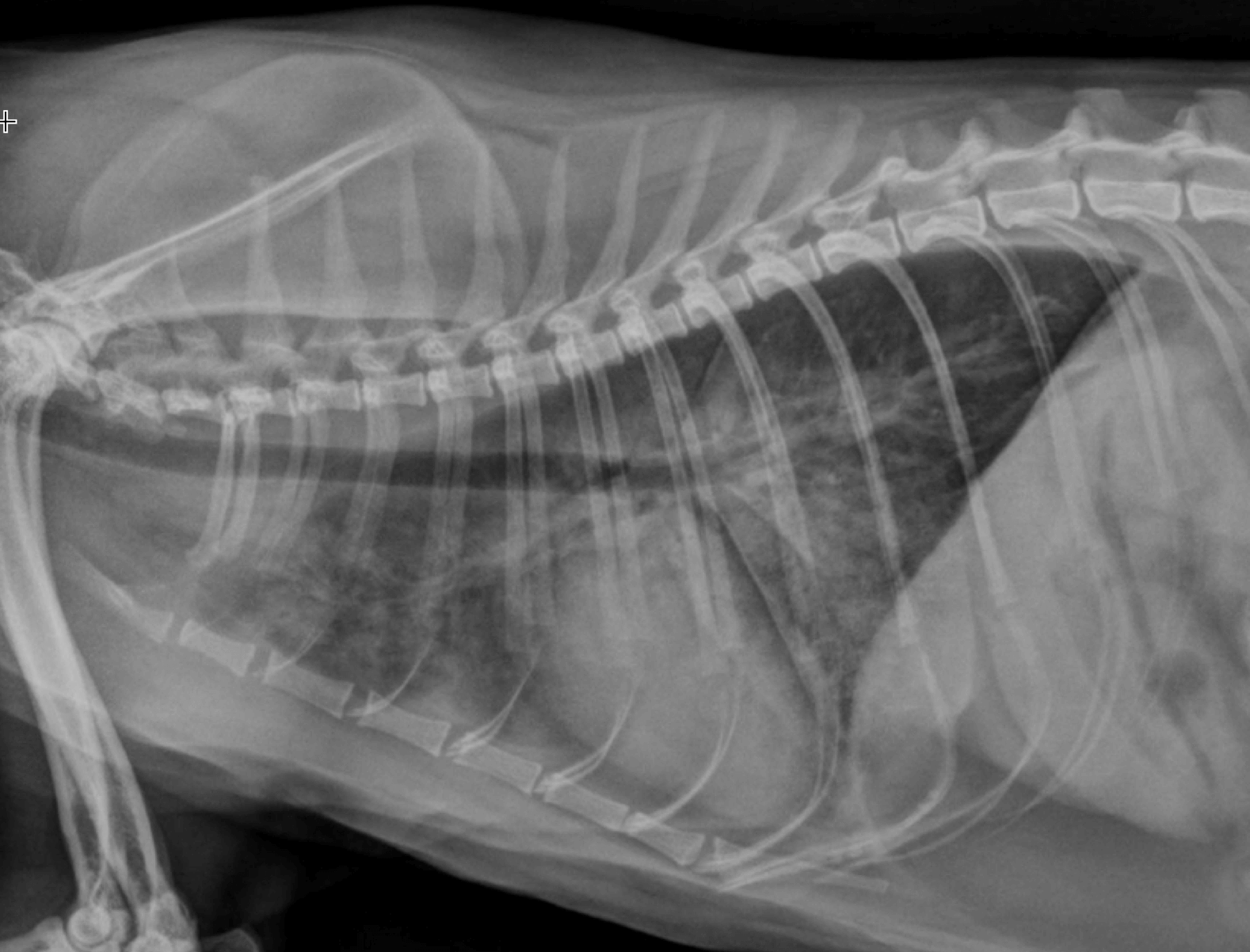
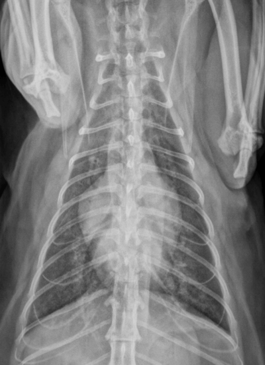
Radiographic interpretation: Generalized cardiomegaly based on increased width on VD view suggestive of atrial enlargement and VHS = 8.8. There is also a moderate to severe mixed interstitial alveolar pattern that is diffuse, but worse ventrally. In addition careful inspection of the pulmonary vessels reveal dilation of the pulmonary veins.
Radiographic diagnosis: Taken together and in light of the presenting complaint and physical examination these finding are highly supportive of a diagnosis of left sided congestive heart failure.
Clinical interpretation/additional case information: For detailed recommendations on management please see the CEG Recommendations “Approach to the dyspneic cat.”
Clinical History
Signalment: 3 year old MC DSH
Clinical history: Patient was presented for respiratory distress. Owner reports that the cat has been acting differently at home for the past two weeks. Rufus appears to be grumpy and is not as active. This morning the owner noticed that Rufus was breathing rapidly and had pale mucous membranes. He usually has a good appetite but he did not eat this morning and is very lethargic. On physical examinationm Rufus was tachypneic and dyspneic. A grade 2/6 sternal systolic heart murmur and gallop heart sounds were heard on auscultation. Rufus received butorphanol and 2mg/kg of furosemide IM and after 30 min of oxygen supplementation the thoracic radiographs were taken.

