Sammy
Saint Bernard
Radiographic Report
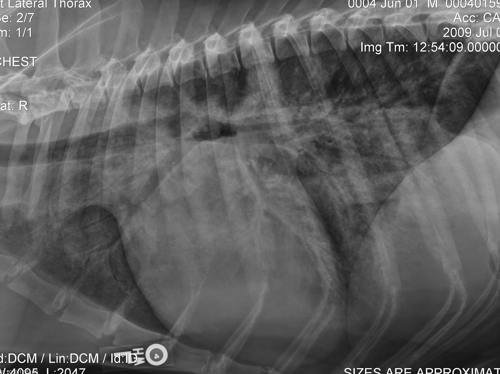
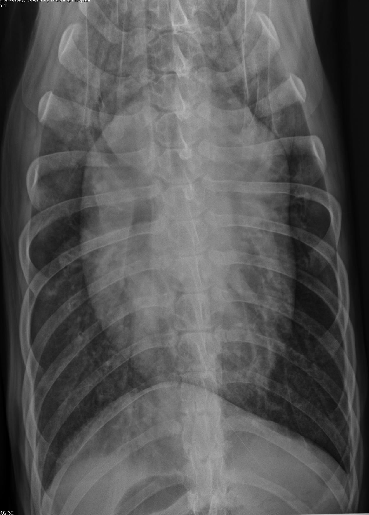
Radiographic interpretation: The heart is subjectively enlarged and the VHS and VLAS are increased. On the VD projection, the left ventricle is enlarged based on marked elongation of the cardiac silhouette and changes in the contour of the heart, showing a left auricular bulge at the 2-3 o’clock position. The lateral projection shows marked left atrial enlargement and prominence of the pulmonary veins (best seen in the cranial lobes). This dog is deep chested, and although the trachea and left lobar bronchus are elevated, the cardiac elongation is less evident than on the VD projection. There are marked increases in the intrapulmonary densities compatible with unstructured interstitial and alveolar pulmonary edema. Acute CHF in large-breed dogs often creates widespread pulmonary edema, including a cranioventral distribution that can be confused with pneumonia. In this patient, there are also marked perihilar increases in lung density that along with left heart enlargement and vascular prominence seal the diagnosis of CHF. Follow-up radiographs (not shown) demonstrated a marked reduction in both cardiac size and lung infiltration after therapy for CHF.
Clinical interpretation/ additional case information: The clinical history is typical of left-sided CHF in dogs with dilated cardiomyopathy. Many of these patients present with a soft cough, and in the absence of a prominent murmur, are treated presumptively for respiratory infection or bronchitis. In this case, a diagnosis of left-sided congestive heart failure is straightforward. Thoracic radiographs are essential to evaluate significant respiratory signs and in this case were diagnostic. Echocardiography indicated a dilated cardiomyopathy phenotype typical of heart disease in giant breeds.
Clinical History
Signalment: 5-year-old MC Saint Bernard dog
Clinical History: Patient was presented with a recent history of coughing and rapid/difficult breathing and now refuses to lay down. Recent laboratory tests were unremarkable, including a negative heartworm test. Sammy has experienced a nearly 20 pound weight loss over the past month. There are no current medications other than heartworm preventative. Physical examination indicated a regular tachycardia with weak arterial pulses; the rhythm was found to be sinus tachycardia on a screening ECG. Cardiac auscultation revealed a loud gallop sound and soft systolic murmur over the mitral valve area. Diffuse pulmonary crackles were identified. After initial treatment with butorphanol, nasal oxygen and one dose of furosemide, thoracic radiographs were obtained.