Shivers
Miniature Poodle
Select Radiograph(s)
Radiographic Report
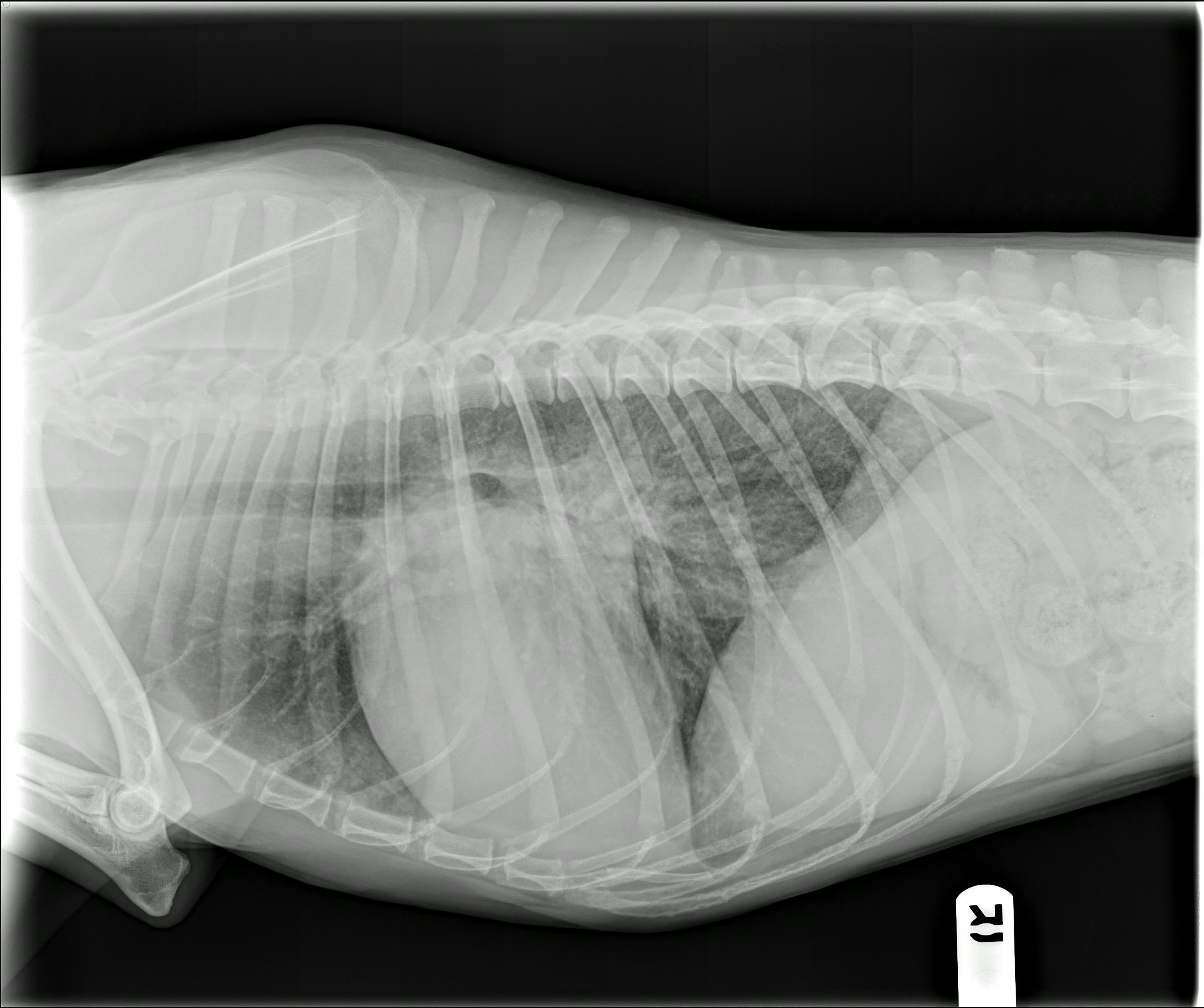
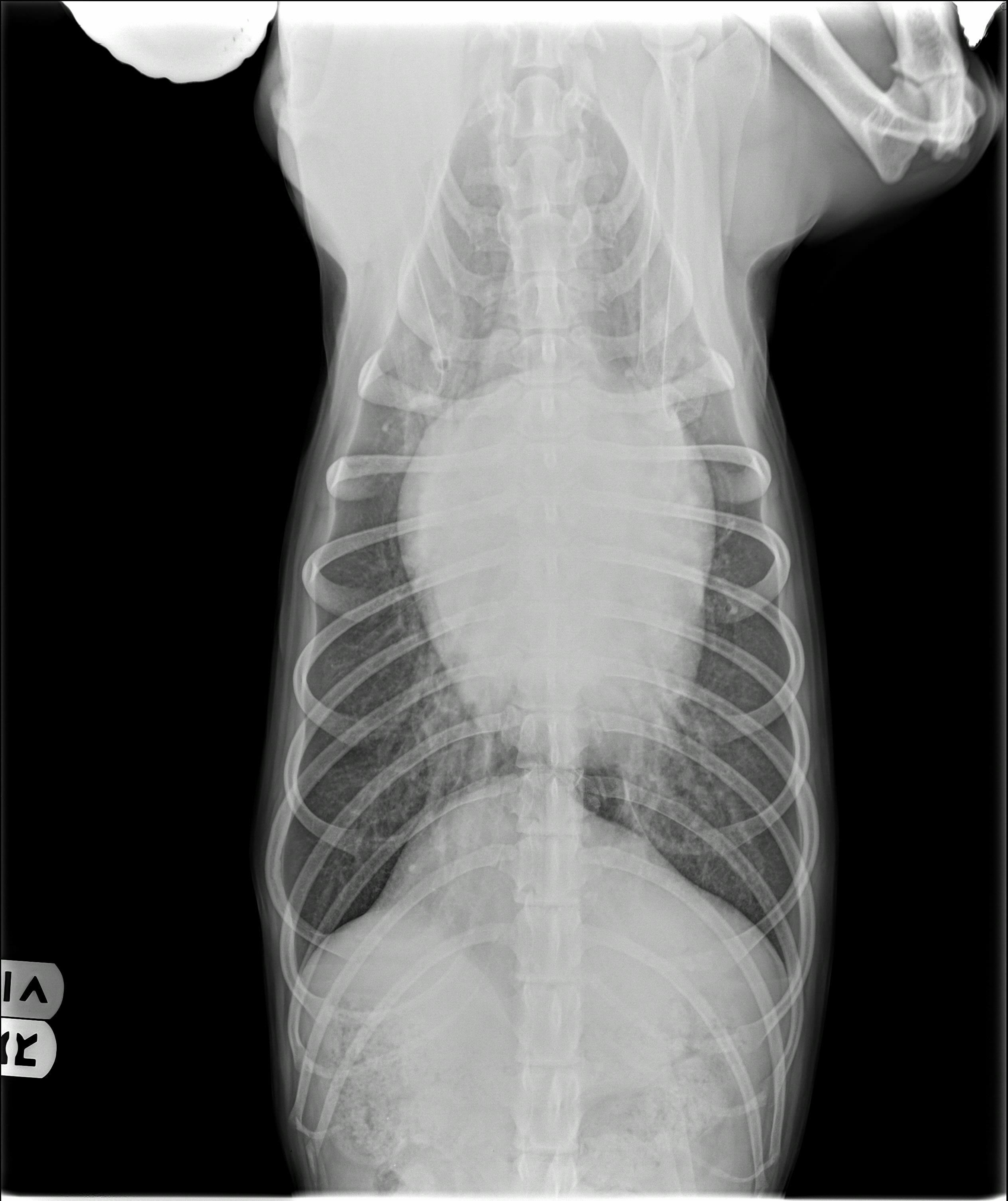
The cardiac silhouette is enlarged with a VHS of 12.4 and a VLAS of 2.7. There is severe left-sided heart enlargement with loss of the caudal waist, indicating left atrial dilation. On the VD view, the cardiac silhouette appears to be wide and somewhat elongated, with a broad left auricular bulge at the 2-3 o’clock position on the cardiac silhouette, consistent with left atrial chamber enlargement. There is a caudal interstitial pattern that is located toward the midline and radiates outward toward the thoracic wall, with the heaviest radiographic density overlaying the left apex of the cardiac silhouette. The pulmonary veins are distended relative to the corresponding arteries.
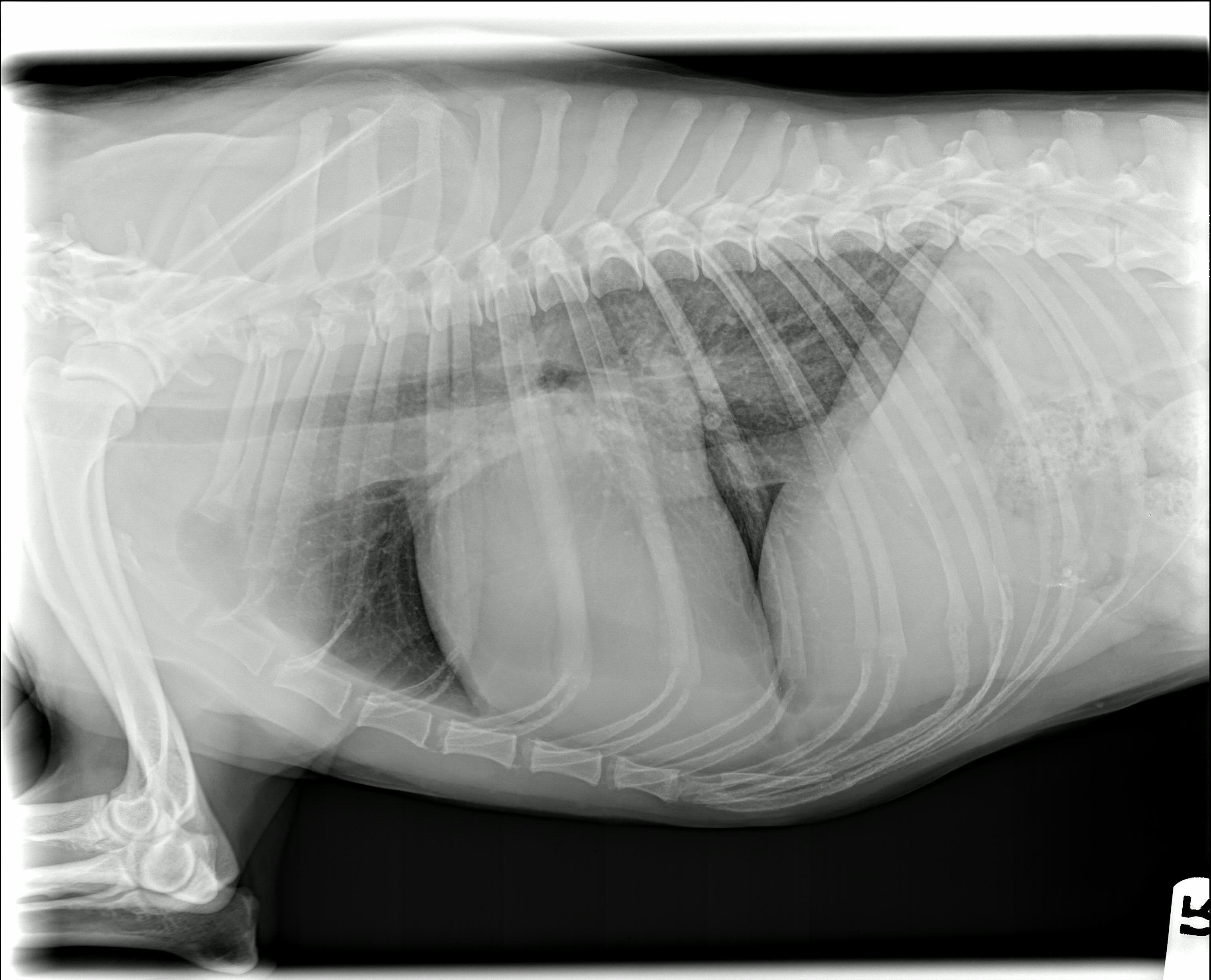
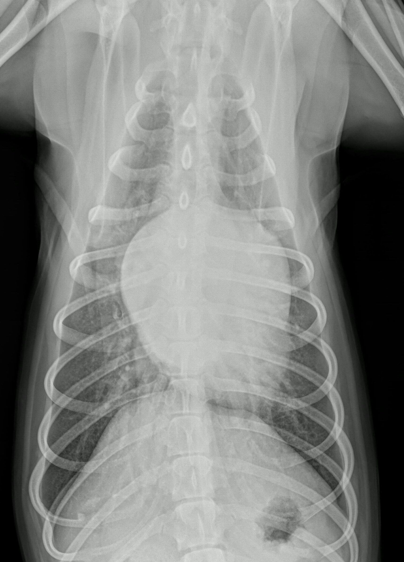
The patient was given a furosemide injection (30mg) subcutaneously and sent home on: Furosemide 20mg tablet every 8 hours by mouth for 3 days then decreased to every 12 hours. Enalapril 5mg tablet every 12 hours by mouth. Pimobendan 2.5mg every 12 hours by mouth. He returned in 7 days for a renal panel and repeat radiographs (see follow up films) which showed improvement in the interstitial pattern, but the pulmonary venous distention had not resolved. Spironolactone 12.5mg every 12 hours was added at that visit.
Clinical History
This is an 11yo male neutered miniature poodle with a primary complaint of rapidly progressive coughing (began 2 weeks ago, but is much more severe over the past 48 hours) accompanied by lethargy and increased respiratory effort. Has a history of a heart murmur, first detected 2 years ago.