Sophie
Bulldog
Select Radiograph(s)
Radiographic Report
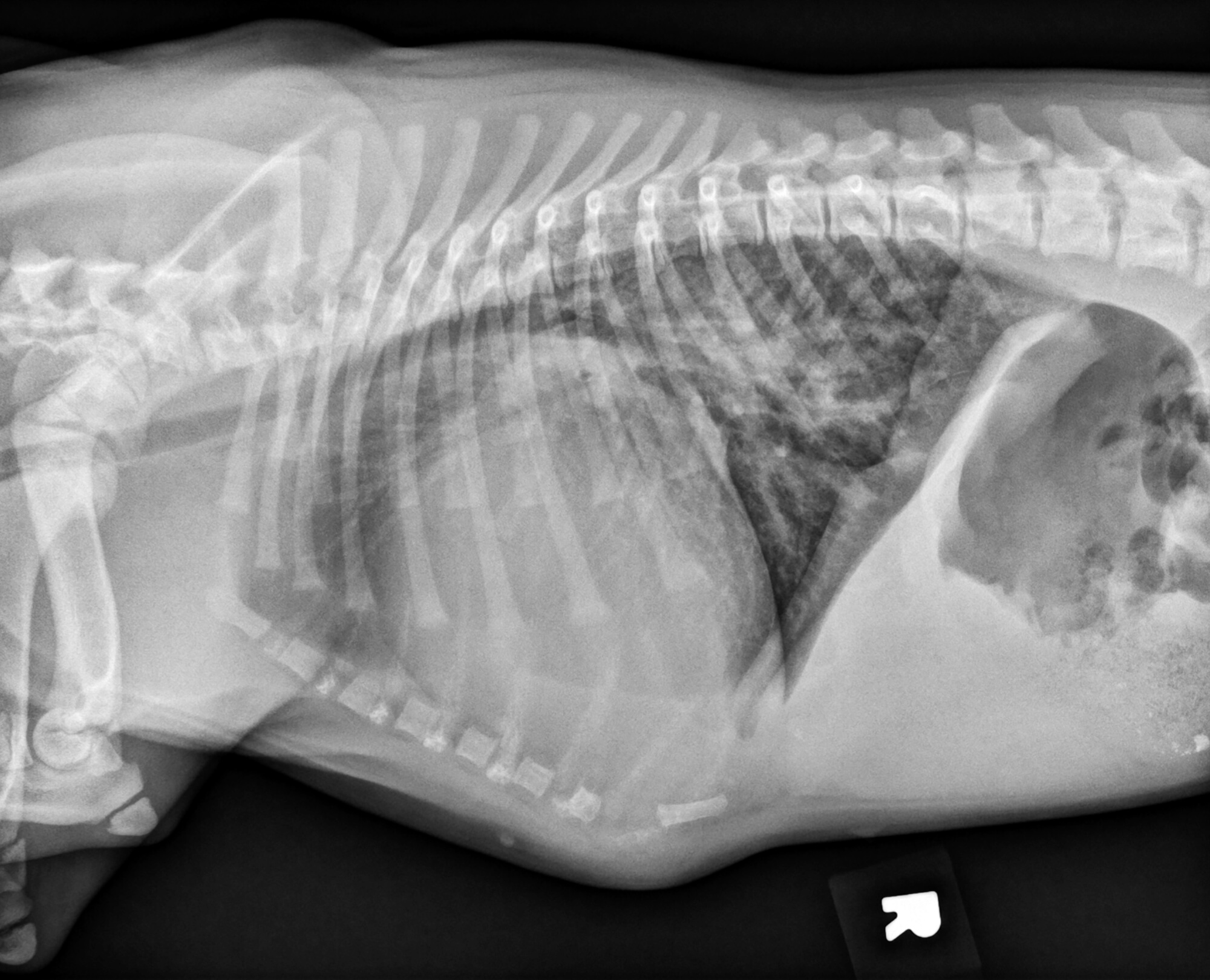
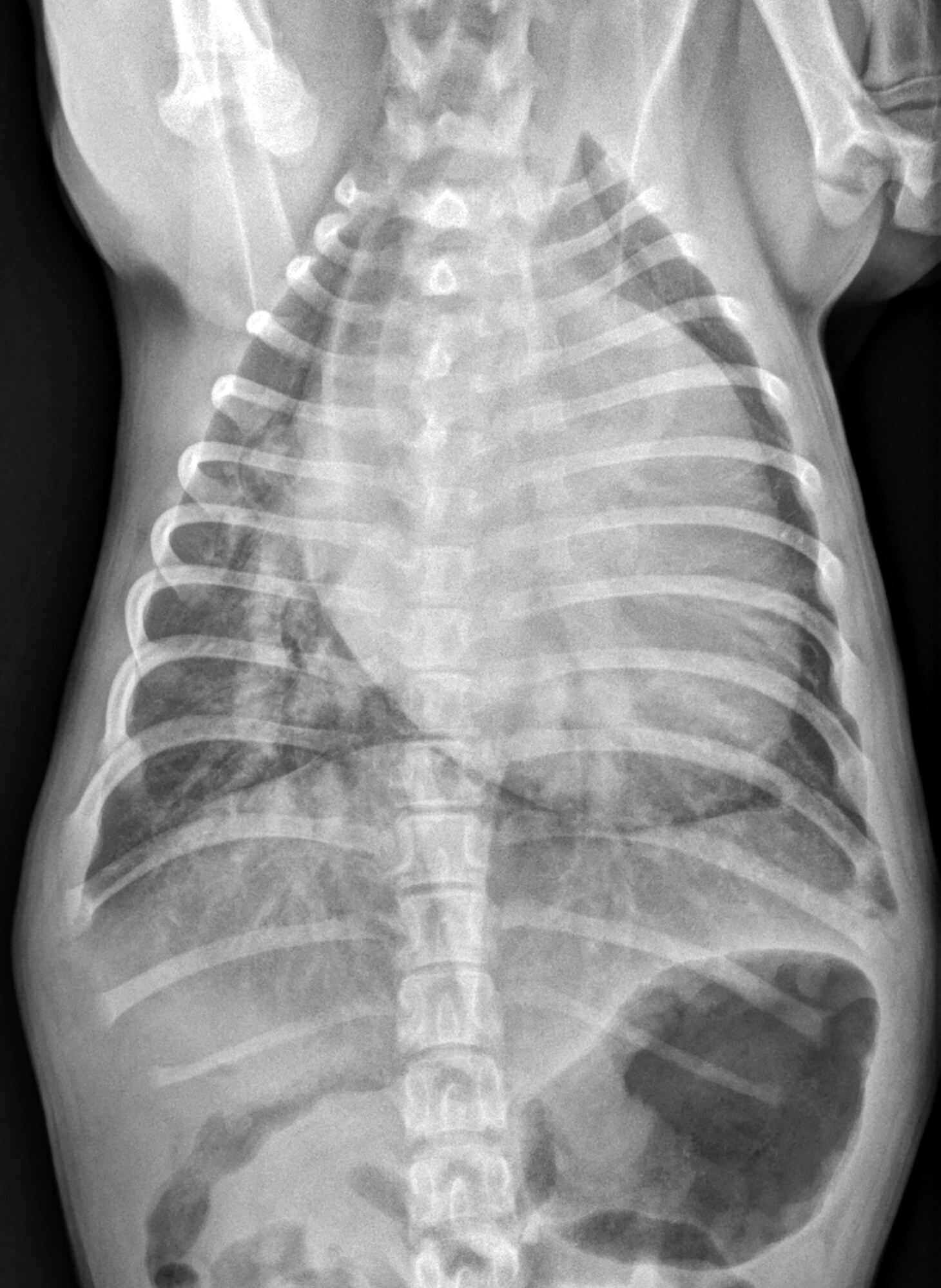
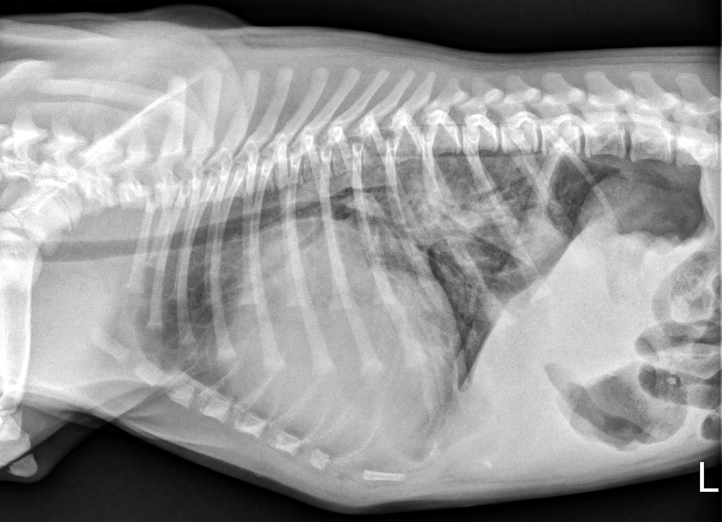
Radiographic interpretation: There is marked generalized cardiomegaly, characterized by an increased height on the lateral projections with mild elevation of the caudal trachea. There is focal enlargement of the left atrium and enlargement of both the pulmonary arteries and veins. There is an unstructured interstitial pattern in the dorsal aspect of the caudal lung lobes and in the perihilar region. The remaining mediastinal structures, pleural space, and diaphragm are within normal limits. There is loss of abdominal serosal detail and multiple open physes, consistent with the age of the patient.
Radiographic diagnosis: The radiographic findings support a diagnosis of decompensated congenital heart disease. Enlargement of both pulmonary arteries and veins are consistent with a left to right shunt, such as a ventricular septal defect or patent ductus arteriosus. The unstructured interstitial pattern is compatible with left sided congestive heart failure.
Clinical interpretation/additional case information: On Sophie’s echocardiogram, subaortic stenosis and a large ventricular septal defect were diagnosed. The left ventricle and left atrium were enlarged. Frequent ventricular ectopy was noted during the cardiac ultrasound. Therapy for the heart failure was advised, but unfortunately the long-term prognosis was grave given two congenital heart defects and development of heart failure at such a young age.
Clinical History
Signalment: 14 week old F American bulldog puppy
Clinical history: This puppy was adopted one month ago, knowing that she had a heart murmur. Since adoption, she had been doing well at home until yesterday when she started to gag/retch. Since then, her condition has worsened as she developed a cough, an elevated respiratory rate, and appeared to vomit, prompting the oner to seek further emergency care. On physical examination, Sophie is tachypneic and dyspneic with increased abdominal effort and harsh lung sounds in all quadrants. A grade V/VI left basilar systolic heart murmur is appreciated, heart rate was 190 bpm, and the femoral pulses were weak. Radiographs were taken to evaluate causes for the dyspnea.