Steinway
Yorkie Mix
Select Radiograph(s)
Radiographic Report
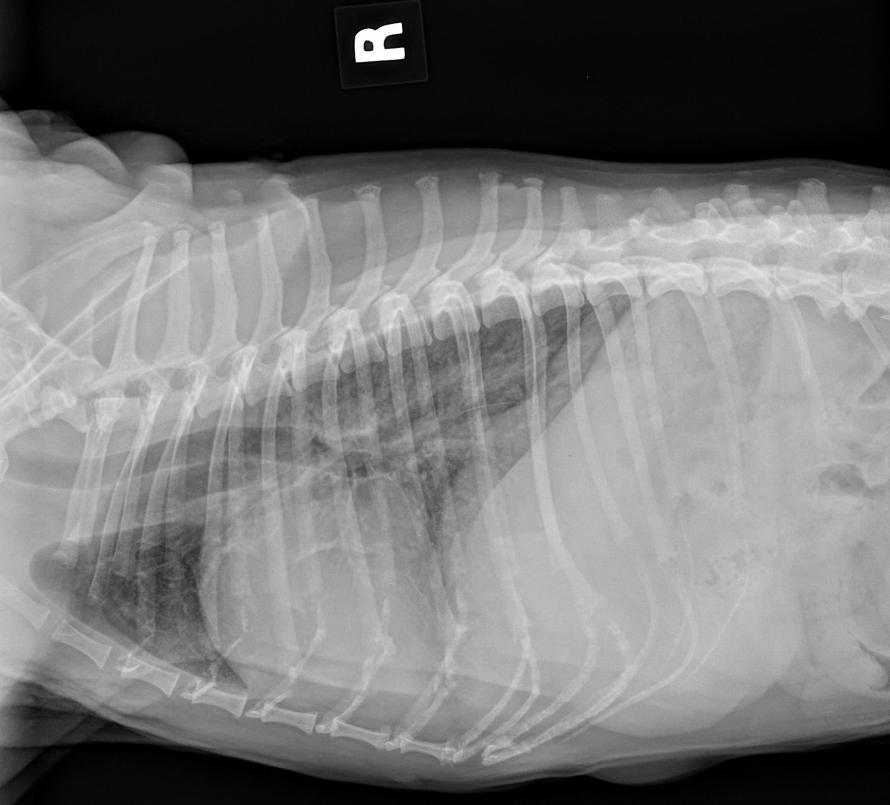
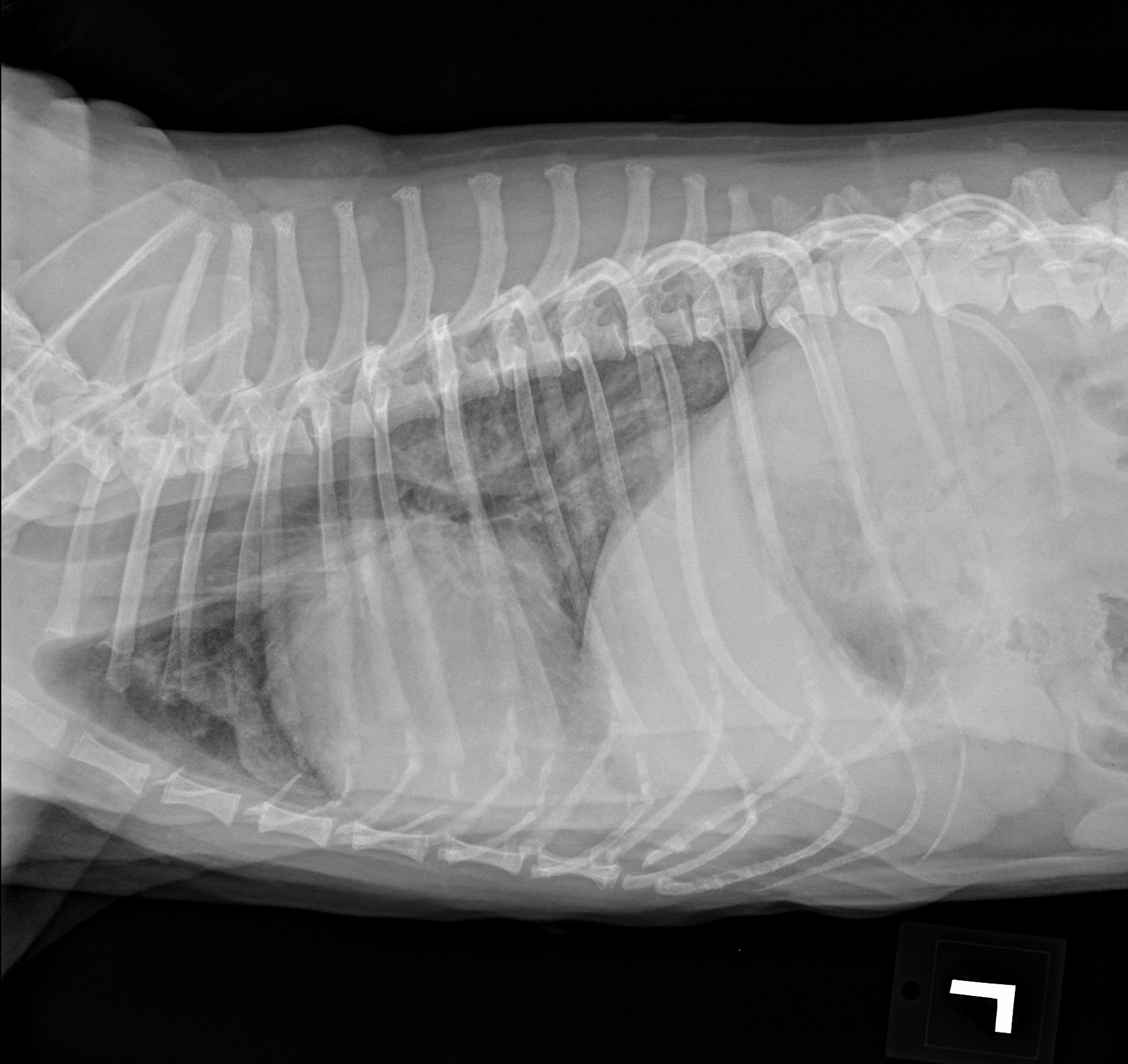
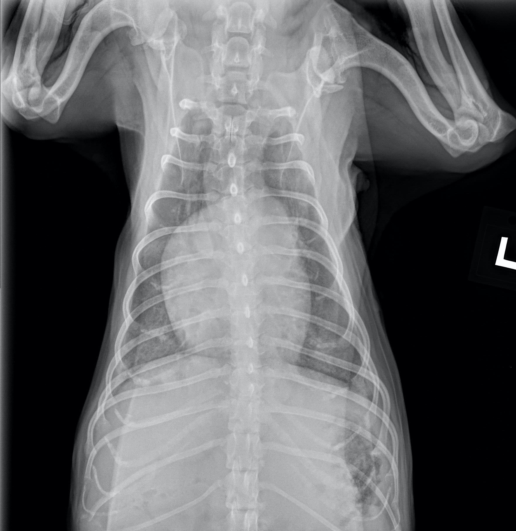
Radiographic interpretation: Patient has suspected enlargement of the right ventricle with a bulge at the 2 O’clock position consistent with dilation of the main pulmonary artery. The right atrium appears mildly dilated. No obvious left heart enlargement is seen. The pulmonary arteries and veins, where visible, do not appear distended. There is a diffuse interstitial pattern consistent with pneumonia or non-cardiogenic pulmonary edema.
Radiographic diagnosis: Findings consistent with pulmonary hypertension and non-cardiogenic pulmonary edema. Consider possible pulmonary thromboembolism.
Clinical interpretation / addition case information: An echocardiogram was performed with the following findings: Eccentric hypertrophy of the right ventricle with dilation of the visible pulmonary arteries. Moderate tricuspid regurgitation with an elevated tricuspid regurgitation velocity consistent with moderate to severe pulmonary hypertension (TR peak pressure gradient=75mmHg). History and diagnostic findings are consistent with acute pulmonary thromboembolism.
Clinical History
Signalment: 13yr old MC Yorkie Mix
Clinical history: Presented to the emergency service for sudden onset respiratory distress. Steinway seemed fine this morning, but around noon, he suddenly starting have tachypnea with mild dyspnea. He is refusing to eat or even lay down. The initial physical examination reveals a new grade III/VI right apical systolic heart murmur and bilateral pulmonary crackles.