Teddy
Pomeranian Mix
Select Radiograph(s)
Radiographic Report
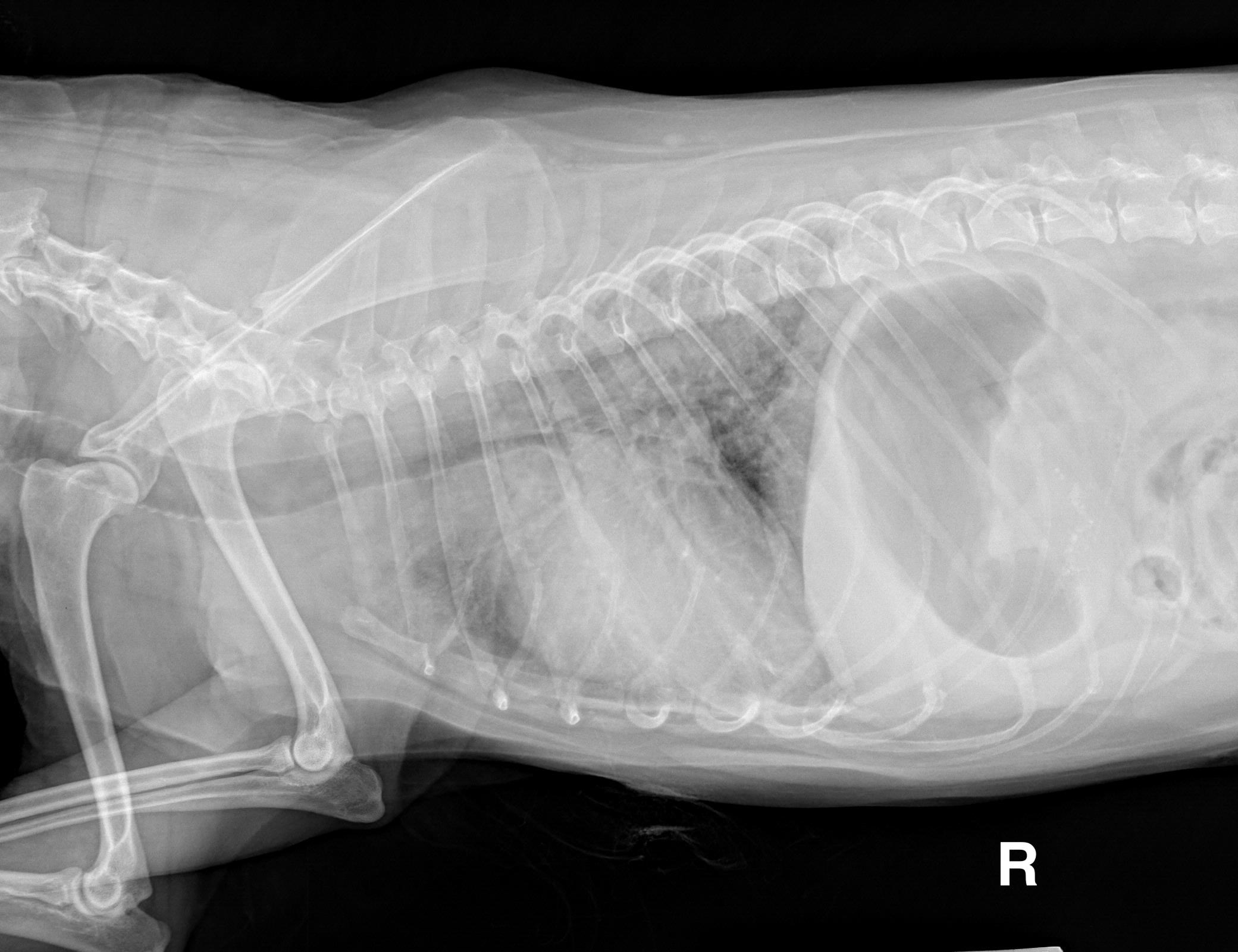
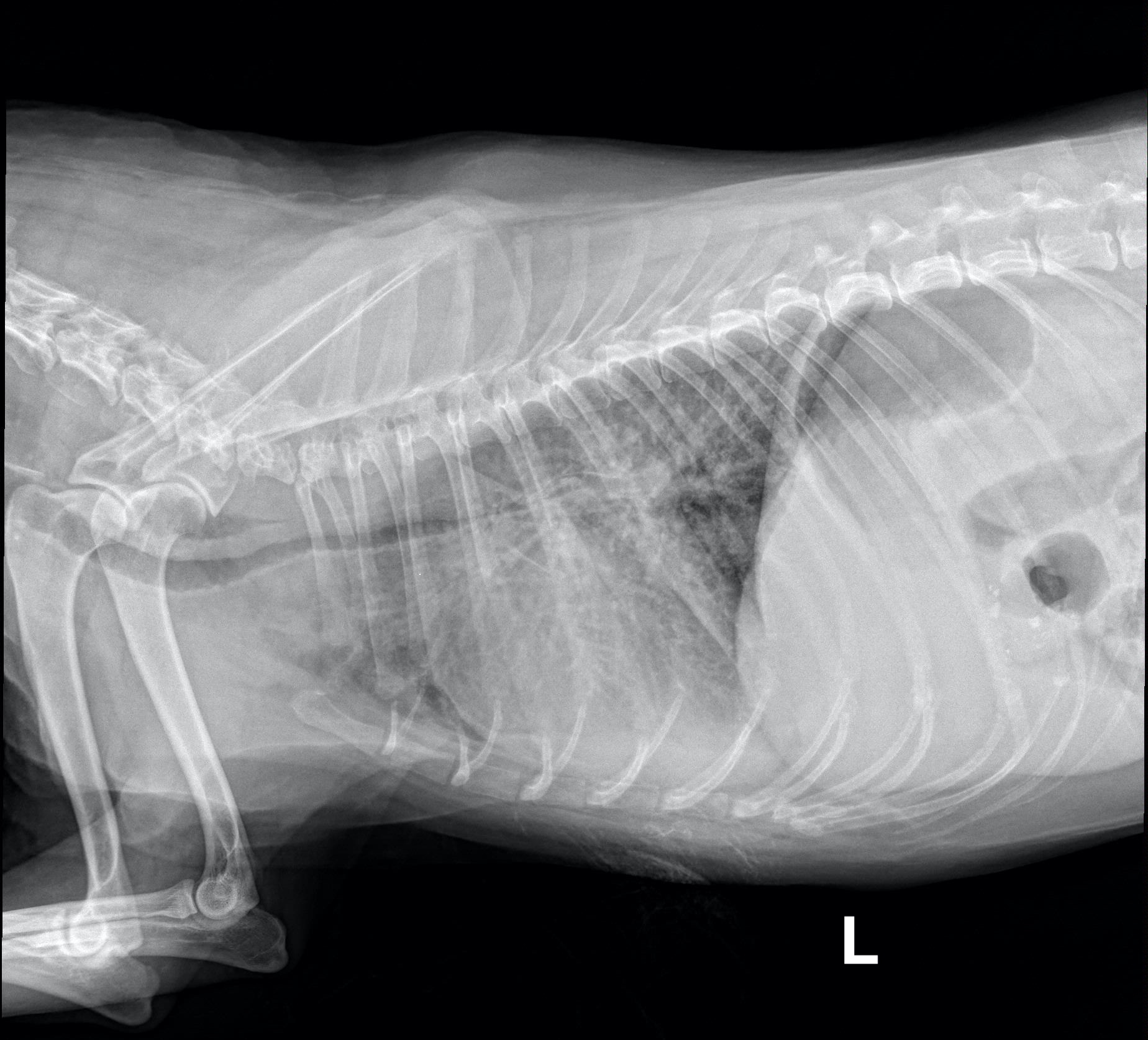
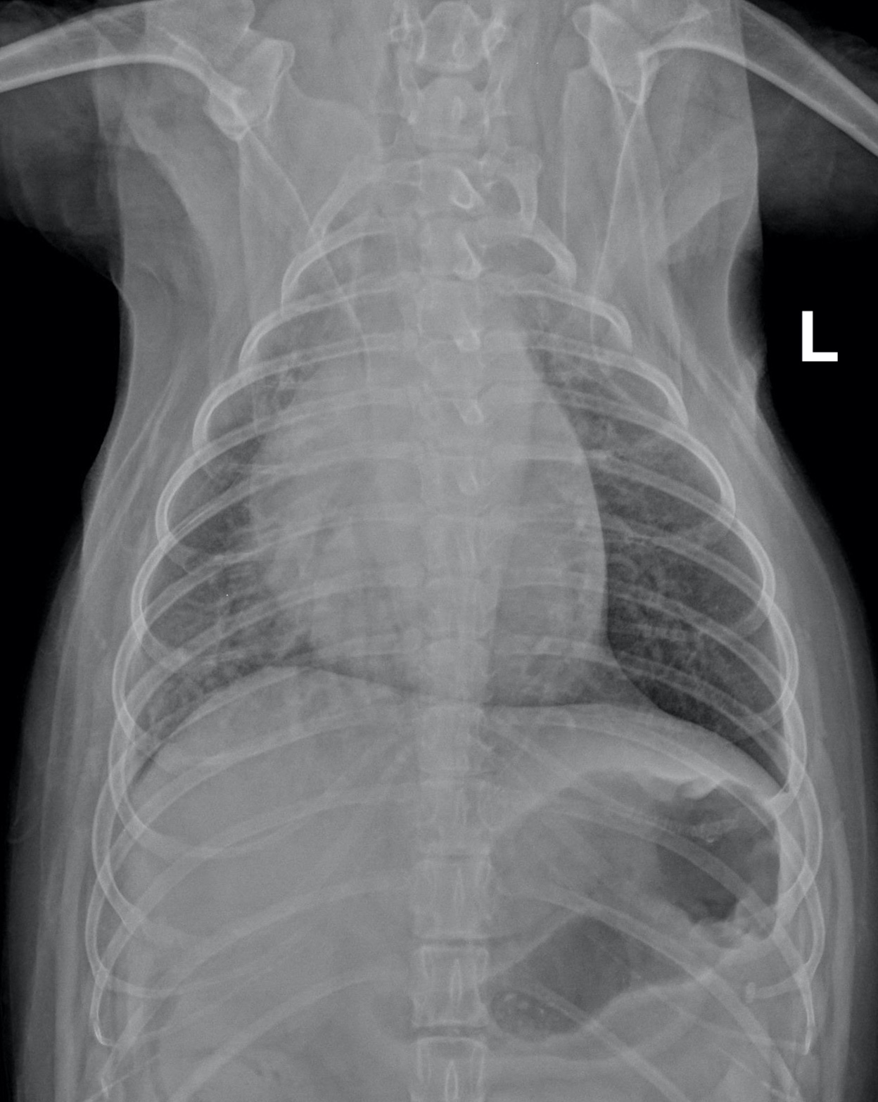
Radiographic interpretation: Suspected enlargement of the left atrium and left ventricle based on a VHS of 11.5 and VLAS of 2.6. The pulmonary arteries and veins, where visible, do not appear distended. There is a diffuse, heavy bronchial to broncho-interstitial pulmonary pattern consistent with chronic lower airway disease. There is also significant narrowing and tortuosity to the thoracic trachea and mainstem bronchi, consistent with tracheal and bronchial collapse. There is no evidence of cardiogenic pulmonary edema.
Clinical interpretation / additional case information: An echocardiogram was performed and moderate myxomatous mitral valve disease was identified as the cause of the murmur, and moderate heart enlargement exceeding EPIC study criteria for pimobendan use was identified. Pimobendan was prescribed in an attempt to delay the onset of congestive heart failure. An appropriate antibiotic and anti-inflammatory dose of steroids were prescribed for the primary pulmonary disease.
Clinical History
Signalment: 12 yr old MC Pomeranian
Clinical history: History of chronic cough for years. About 18 months ago, a Grade III/VI left apical systolic murmur was found during evaluation for worsening cough, but no cardiac treatment was given. The patient was presented today for an acute exacerbation of the chronic cough. The patient has a sleeping breathing rate of 24 breaths/min at home. The initial physical examination revealed a loud heart murmur (grade IV/VI left apical systolic), a honking cough and increased bronchovesicular lung sounds bilaterally. A CBC reveals a normal WBC count.