Tide
Newfoundland
Select Radiograph(s)
Radiographic Report
Radiographic interpretation:
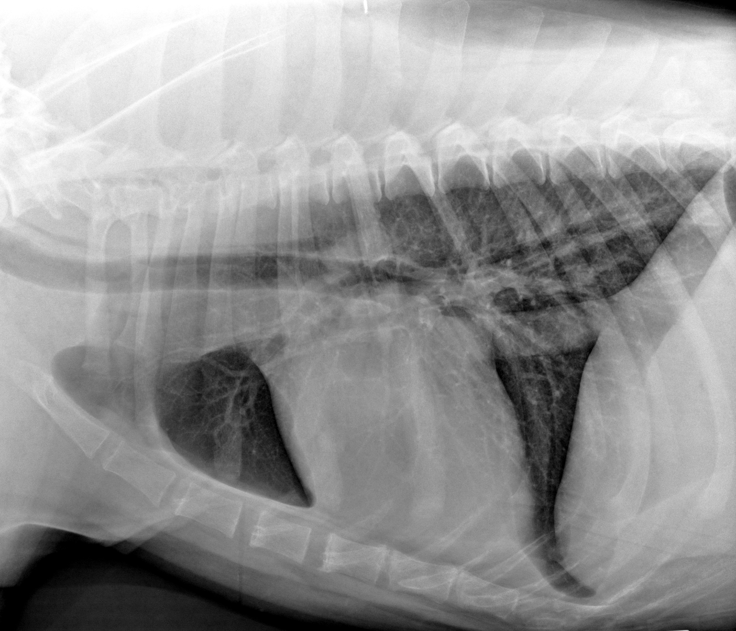
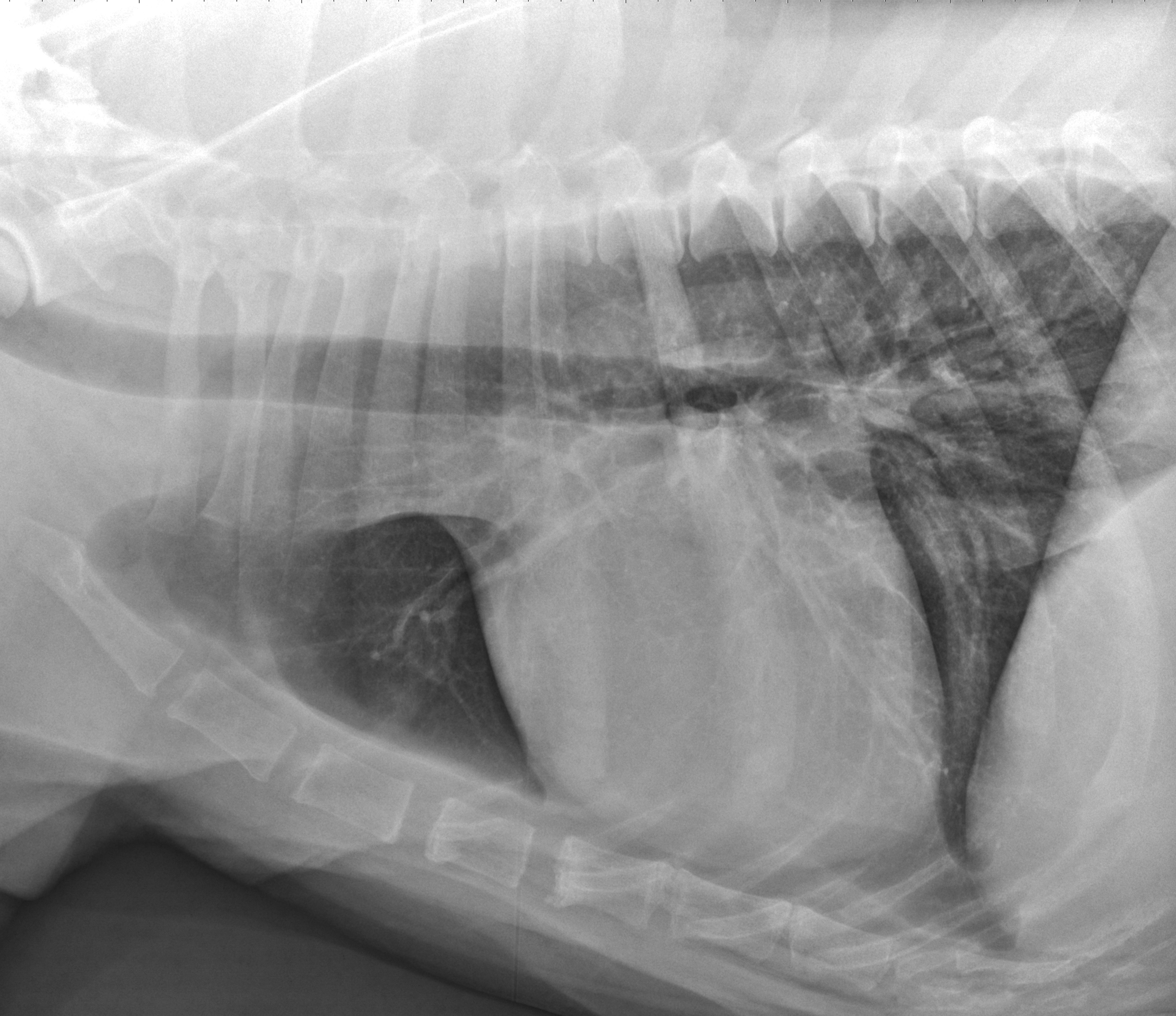
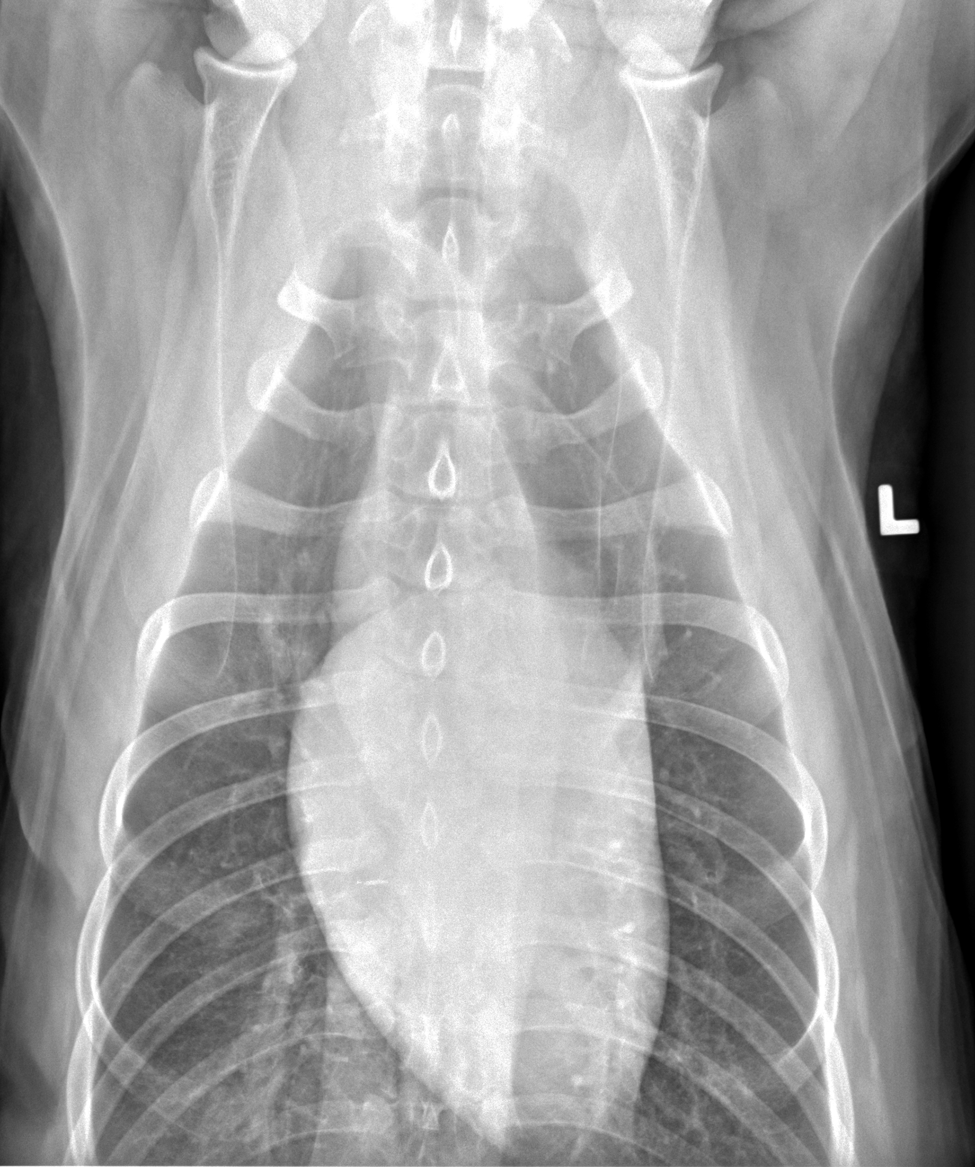
The cardiac silhouette is normal in size and shape. There is small amount of gas collecting within the esophagus. The pulmonary parenchyma, pulmonary vasculature and pleura are normal. There is no evidence of pulmonary metastases. There is mild osteoarthritic changes of the facet joints of the caudal thoracic and cranial lumbar vertebrae. The remainder of the extrathoracic osseus and soft tissue structures are normal.
Clinical interpretation/ additional case information:
There is no intrathoracic pathology to explain the restless signs nor a clear explanation for the atrial fibrillation. In giant breeds of dog (Newfoundland, Irish wolfhound, great Dane, etc), atrial fibrillation may develop in a structurally normal heart – similar to what occurs in horses. Echocardiography in this dog was performed and found normal chamber sizes, normal systolic function, no valvular regurgitation, and no congenital heart defects. As such, the diagnosis was lone atrial fibrillation, meaning development of the rhythm disturbance in the absence of structural heart disease. Treatment options include rhythm control (electrical cardioversion to restore sinus rhythm) or cardioprotective medications with rate control. A rate control strategy means that the patient remains in atrial fibrillation, but the ventricular response rate is monitored and rate modulating medications prescribed if the rate is high. This dog received carvedilol for cardioprotection and for rate control. He has done well with his heart disease since initial diagnosis. The cause of the nonspecific clinical signs is unclear, though may reflect discomfort from acute onset of the arrhythmia.
Clinical History
Signalment: 5-year-old, MC, Newfoundland dog
Clinical History
This dog has a 3-day history of restlessness and what the client perceived as pain or discomfort. There is no history of other relevant disease and he has been owned by the client since 10 weeks of age. Physical examination detected an irregular heart rhythm, no heart murmur, normal lung sounds, and no other apparent cause for the clinical signs. The irregular heart rhythm had a rate of 130 beats per minute and ECG showed atrial fibrillation. Radiographs were taken to evaluate underlying heart size and for the presence of any intrathoracic disease as a cause of the restlessness.


