Tigger
DSH
Radiographic Report
Radiographic interpretation:
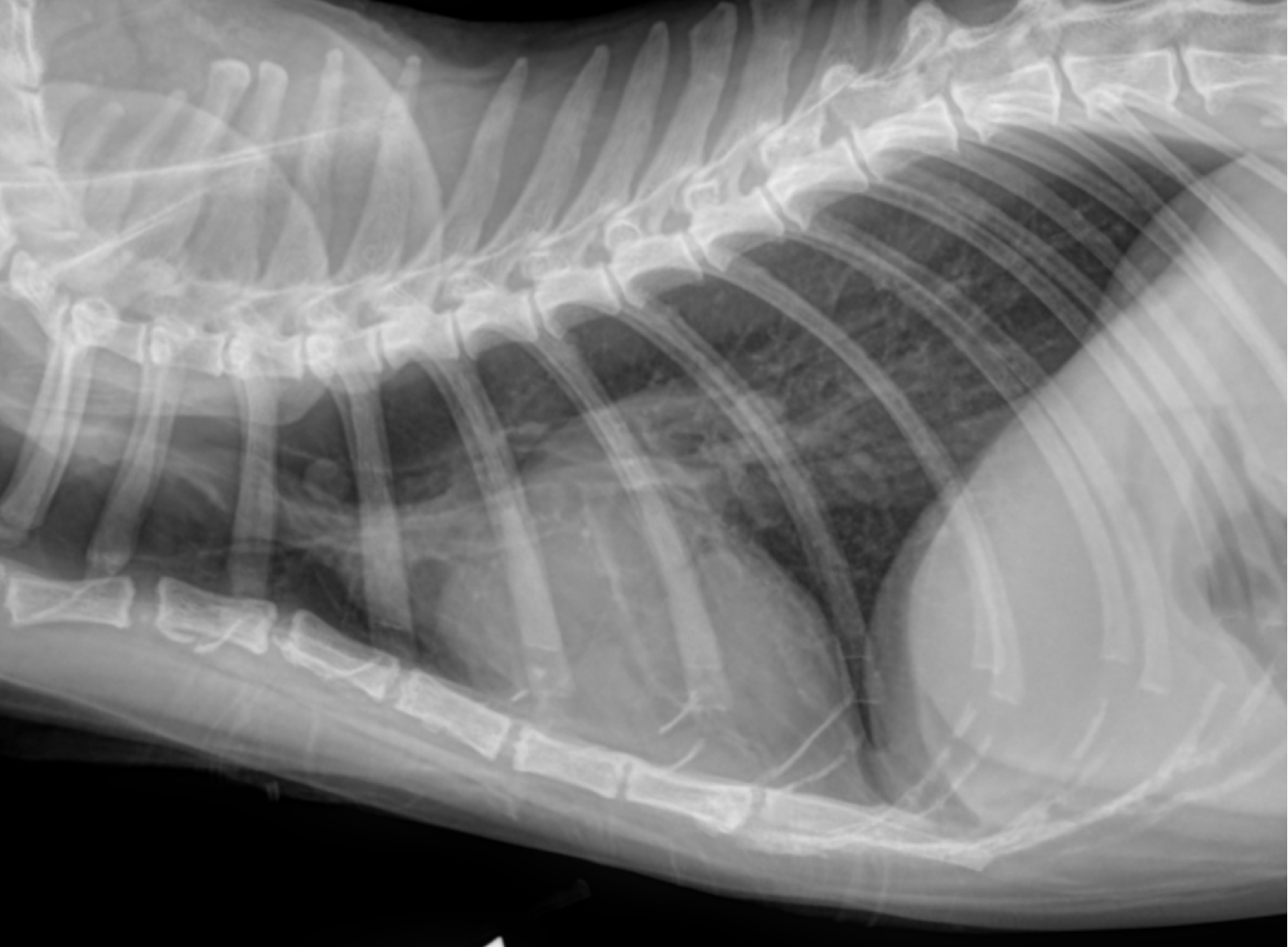
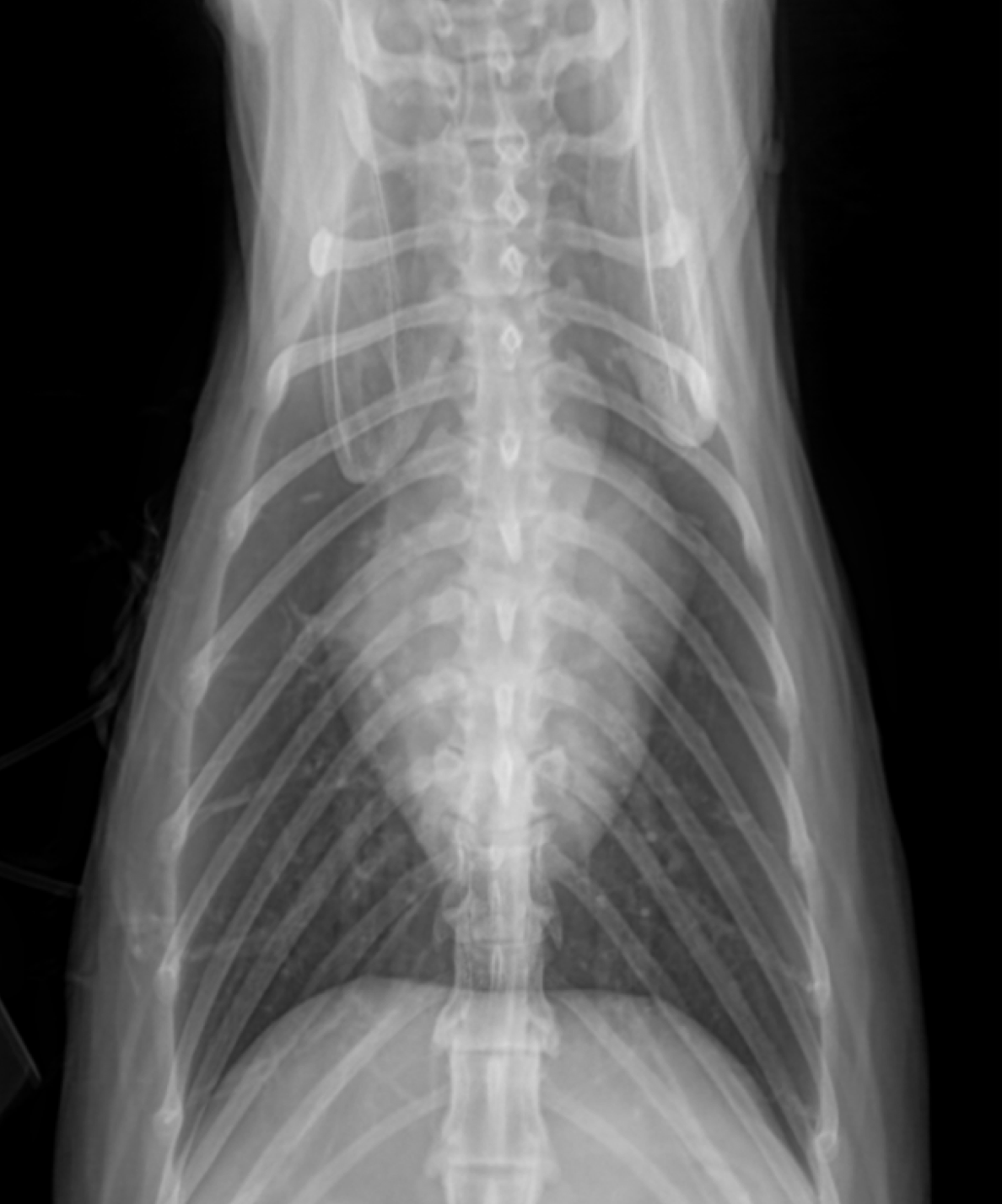
The cardiac silhouette is enlarged with a VHS of 8.8 and a classic valentine heart appearance on the VD. There is no evidence of pulmonary edema or pleural effusion, but the vasculature is prominent, especially the caudal vessels on the lateral view. Incidental pulmonary nodules are present in the 3rd intercostal space and also over the 7th rib.
Radiographic Diagnosis: Cardiac enlargement (especially left atrial) with prominent vasculature, but no signs of CHF.
Clinical interpretation / additional case information: No evidence for CHF is identified. The enlarged heart and the increased vasculature seen on the lateral views suggests that heart disease is present and hemodynamically significant, but CHF has not yet developed.
Clinical History
Signalment: 19 year old MC Domestic Shorthair
Clinical History: Presented for vomiting x 1 week with acute progression. Patient is lethargic and has stopped eating. There is a 3/6 left sternal systolic murmur. Blood work identifies significant azotemia (BUN 102, crea 2.5).