Tilly
Labrador Retriever
Radiographic Report
Radiographic interpretation:Later
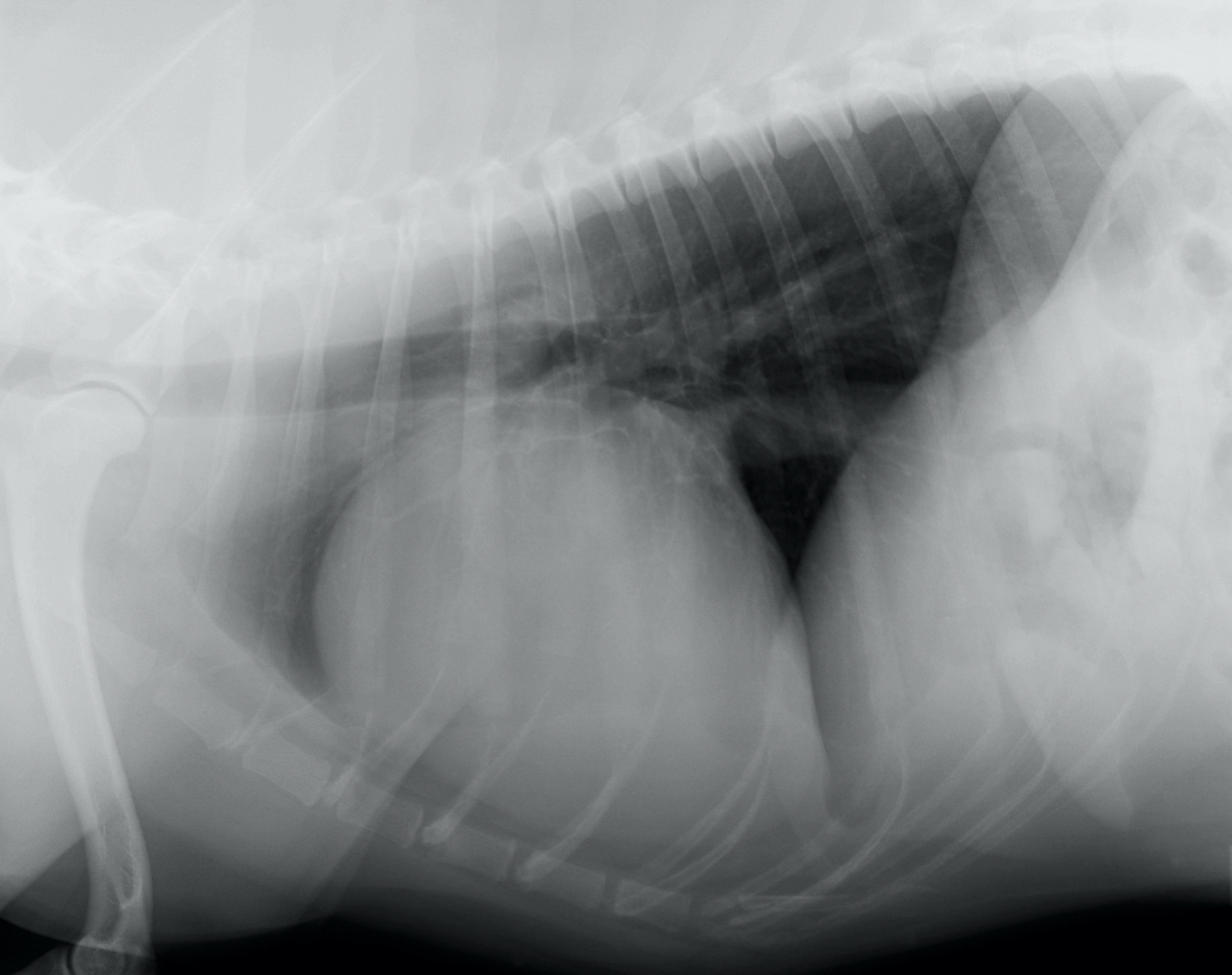
Lateral radiograph: Cardiomegaly is present with a VHS of 12.4. The carina is displaced dorsally, a finding often produced by left ventricular enlargement. However in this case, the more prominent finding is increased sternal contact and lifting of the apex off the sternum suggestive of right ventricular enlargement. Therefore, the deviation of the carina may instead be related to right ventricular enlargement. The caudal and cranial cardiac silhouettes are normal. The caudal vena cava is distended.
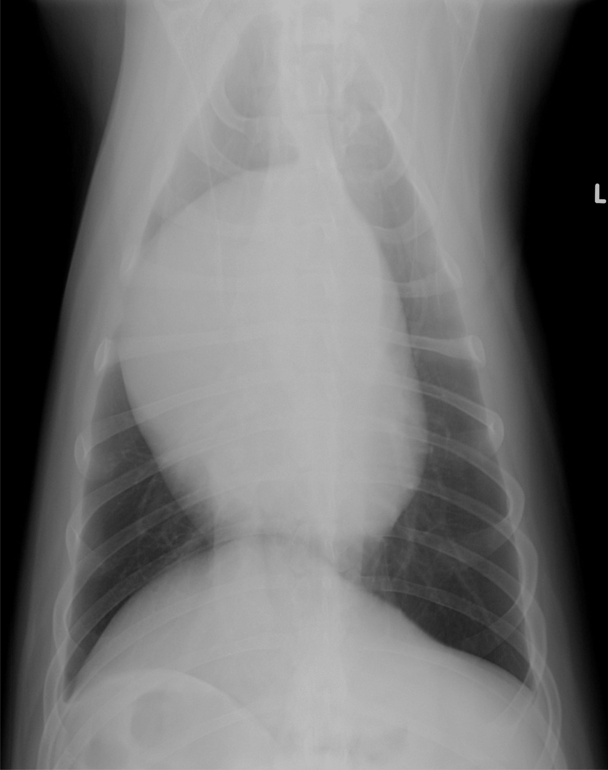
Ventrodorsal radiograph: The border of the left heart is straight while the right ventricle and right atrium appear enlarged (6 to 12 o’clock). There is no evidence of left atrial or left auricular enlargement. The aortic arch and main pulmonary artery appear normal.
Radiographic diagnosis:Radiographic interpretation: Right ventricular and right atrial enlargement with distension of the caudal vena cava, notable in the VD projection.
Additional information: Echocardiography revealed tricuspid valve dysplasia (congenital malformation of the tricuspid valve) with tricuspid regurgitation.
Clinical History
Signalment: 3 year old FS Labrador
Clinical history: Tilly was presented for a routine preventative health examination after relocating to your area 7 months ago. The owners report she is asymptomatic at home. Auscultation reveals a grade III/VI systolic right-sided murmur with no audible arrhythmias. There is jugular pulsation in the lower 1/2 of the neck. Femoral pulses are strong and lung sounds are normal.